127 posters, 10 topics, 20 sessions, 127 authors
ePostersLive by SciGen Technologies S.A. All rights reserved.
UCSF 2024 Inquiry Symposium
Conference ePosters

P077
Race/ethnicity and socioeconomic-based analysis of vascular alterations on optical coherence tomography angiography (OCTA) in patients with diabetes
Poster Presenter
Authors
Part of Topic
Clinical & Translational Sciences
Race/ethnicity and socioeconomic-based analysis of vascular alterations on optical coherence tomography angiography (OCTA) in patients with diabetes
Abu T Taha, Yi Stephanie Zhang, Isabel Thompson, Jeremy D Keenan, Aunoy Poddar, John Dickson, Ricardo Lamy, Jay M Stewart
Purpose
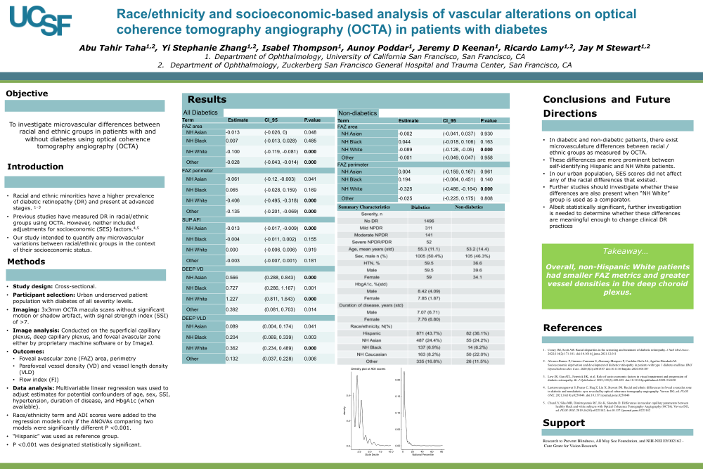
Racial and ethnic minorities have a higher prevalence of diabetic retinopathy (DR) and present at advanced stages.1–3 Two studies have measured DR in racial/ethnic groups using OCTA. However, neither included adjustments for socioeconomic (SES) factors.4,5 We performed a retrospective observational study using optical coherence tomography angiography (OCTA) to investigate microvascular differences between racial/ethnic groups and their respective SES scores from the area deprivation index (ADI). Patients with diabetes were grouped into: no DR, mild to severe non-proliferative diabetic retinopathy (NPDR), and proliferative diabetic retinopathy (PDR).
Methods:
We obtained 3x3mm macular OCTA scans from patients without other ocular comorbidities or prior DR treatments. One eye per patient was included in the analysis. Parameters analyzed included foveal avascular zone (FAZ), superficial capillary plexus (SCP) and deep capillary plexus (DCP) for parafoveal vessel density (VD), vessel length density (VLD), and flow index (FI). SES was measured using patients’ addresses and ADI. Multivariable linear regression was used to adjust estimates for potential confounders of age, SS, hypertension, gender, duration of disease, and HbgA1C.
Results:
227 controls and 1993 diabetics were included in the study. Racial breakdown was 43% Hispanic, 24.4% NH Asian, 6.8% NH Black, 9.6% NH Caucasians, and 16.3% Other. In all comparisons, Hispanic group was used as the referent. Among controls, Caucasian patients had smaller FAZ area (ß = -0.089, P < 0.001) and FAZ perimeter (ß = -0.325, P < 0.001). Among all diabetics combined, Caucasians had smaller FAZ area (ß = -0.100, P < 0.001) and FAZ perimeter (ß = -0.406, P < 0.001), but higher deep VD (ß = 1.227, P < 0.001) and deep VLD (ß = 0.362, P < 0.001). Asian participants also had higher deep VD (ß = 0.566, P < 0.001).
Additionally, “Other” had smaller FAZ area (ß = -0.028, P < 0.001) and FAZ perimeter (ß = - 0.135, P < 0.001). Lastly, the addition of SES scores both at a national and state level, did not modify any of the racial regression models significantly.
Conclusions/Discussion
Our findings from a large county population suggest that at baseline, there are differences in the microvascular alterations between racial/ethnic groups that cannot be explained by their SES. Albeit statistically significant, further investigation is needed to determine whether these differences are meaningful enough to change clinical DR practices.
- Coney JM, Scott AW. Racial disparities in the screening and treatment of diabetic retinopathy. J Natl Med Assoc. 2022;114(2):171-181. doi:10.1016/j.jnma.2021.12.011
- Alvarez-Ramos P, Jimenez-Carmona S, Alemany-Marquez P, Cordoba-Doña JA, Aguilar- Diosdado M. Socioeconomic deprivation and development of diabetic retinopathy in patients with type 1 diabetes mellitus. BMJ Open Diabetes Res Care. 2020;8(2):e001387. doi:10.1136/bmjdrc-2020-001387
- Low JR, Gan ATL, Fenwick EK, et al. Role of socio-economic factors in visual impairment and progression of diabetic retinopathy. Br J Ophthalmol. 2021;105(3):420-425. doi:10.1136/bjophthalmol-2020-316430
- Laotaweerungsawat S, Psaras C, Haq Z, Liu X, Stewart JM. Racial and ethnic differences in foveal avascular zone in diabetic and nondiabetic eyes revealed by optical coherence tomography angiography. Vavvas DG, ed. PLOS ONE. 2021;16(10):e0258848. doi:10.1371/journal.pone.0258848
- Chun LY, Silas MR, Dimitroyannis RC, Ho K, Skondra D. Differences in macular capillary parameters between healthy black and white subjects with Optical Coherence Tomography Angiography (OCTA). Vavvas DG, ed. PLOS ONE. 2019;14(10):e0223142. doi:10.1371/journal.pone.0223142