127 posters, 10 topics, 20 sessions, 127 authors
ePostersLive by SciGen Technologies S.A. All rights reserved.
UCSF 2024 Inquiry Symposium
Conference ePosters

P060
Responses in Non-Small Cell Lung Cancer (NSCLC) treated with Neoadjuvant Pembrolizumab and Radiation Therapy (SRT)
Poster Presenter
Authors
Part of Topic
Clinical & Translational Sciences
Title: Single Cell Dissection of Immune Responses in Non-Small Cell Lung Cancer treated with Neoadjuvant Pembrolizumab and Radiation Therapy (SRT).
Authors: Anna Crosetti, Karen Law, Lawrence Fong, MD
Introduction: Pembrolizumab checkpoint inhibition (CI) has transformed the treatment landscape for NSCLC. The effect of neoadjuvant CI with the cell ablative effects of radiation are not well described and may promote a pro-inflammatory antitumor environment, as seen in numerous mouse models. Single cell RNA sequencing (scRNAseq) offers high resolution insight into how individual cells respond to these therapies.
Hypothesis: We hypothesized that the combination of CI and SRT would increase t-cell infiltration into the tumor microenvironment as well as increase t-cell heterogeneity.
Aims: Our study aim was to broadly define the immunologic response to CI and SRT at the single-cell level.
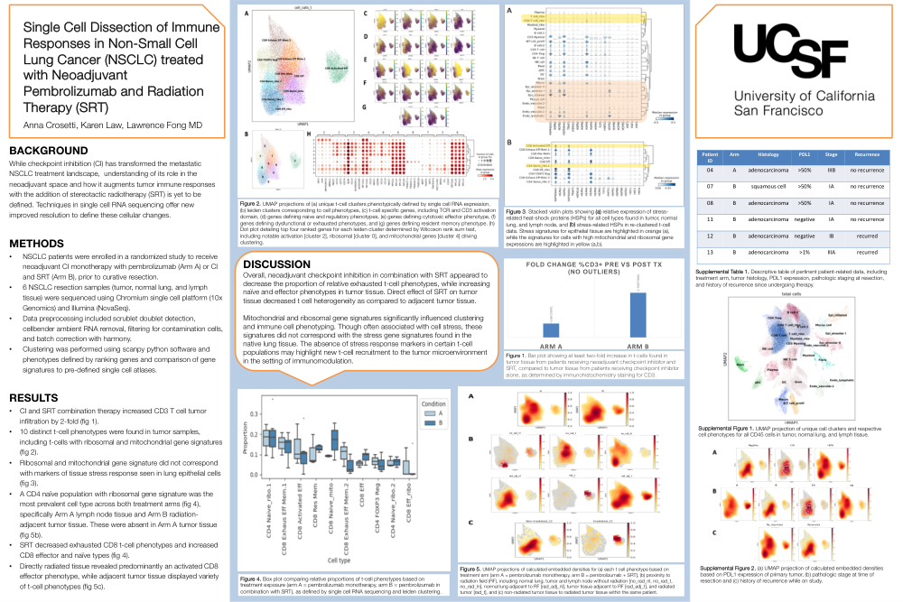
Methods: 12 patients with resectable NSCLC were randomized to 2 cycles of CI monotherapy (Arm A) or in combination with SRT (Arm B) before surgical resection. Tumor, normal lung, and lymph node tissue for 6 participants were sequenced using the Chromium single cell platform (10x Genomics) and Illumina (NovaSeq). Preprocessing included, scrublet doublet detection, cellbender ambient RNA removal, harmony batch correction, and mitochondrial, RBC, and platelet contaminant filtering. Cell clustering, phenotyping, and cell proportion comparisons were performed using scanpy and scCODA python software.
Results: Overall, tumor tissue exposed to CI and SRT displayed a 2-fold increase in t-cell infiltration, based on immunohistochemical staining for t-cell CD3. Analysis via scRNAseq revealed 10 distinct t-cell phenotypes, including cells with high mitochondrial and ribosomal gene expression. These cells lacked stress response gene signatures seen in tumor-adjacent epithelial tissue. CI with SRT decreased the proportion of exhausted CD8 t-cell phenotypes and increased CD8 effector and naïve cell types. Directly irradiated tissue revealed an activated CD8 effector phenotype, while adjacent non-irradiated tumor tissue displayed more variety of t-cell phenotypes.
Conclusions: CI in combination with SRT increased t-cell infiltration in tumor tissue, increased CD4 and CD8 naïve cell populations, while decreasing exhaustive cell states that may promote tumor growth. These populations revealed absent stress signatures seen in adjacent lung tissue, suggesting new infiltration following tissue damage and immune augmentation with CI.F