127 posters, 10 topics, 20 sessions, 127 authors
ePostersLive by SciGen Technologies S.A. All rights reserved.
UCSF 2024 Inquiry Symposium
Conference ePosters

P028
Auto-antibody discovery in primary biliary cholangitis (PBC) using phage-immunoprecipitation sequencing (PhIP-seq)
Poster Presenter
Authors
Part of Topic
Clinical & Translational Sciences
Title: Auto-antibody discovery in primary biliary cholangitis (PBC) using phageimmunoprecipitation sequencing (PhIP-seq)
Introduction: Primary biliary cholangitis (PBC) is an autoimmune liver disease affecting the small bile ducts. Although bile duct damage in PBC is thought to be mediated by immune cells, immunosuppressive treatments have shown little promise. This contradiction indicates our understanding of PBC pathophysiology remains incomplete. Since the current PBC diagnostic markers (ie. anti-mitochondrial antibodies or AMA and anti-nuclear antibodies or ANA) are highly sensitive, this project’s goal is not to establish new diagnostics. Instead, we focus on characterizing the auto-antibody landscape in PBC to further understand its disease mechanism. For auto-antibody determination, we will be utilizing a phage-immunoprecipitation sequencing (PhIP-seq) approach.
Hypothesis: We hypothesize that certain auto-antibodies will be associated with PBC when compared to healthy patients.
Aim: Characterize the auto-antibody landscape in serum of patients with PBC and compare it to healthy patients controls using PhIP-seq. Validate hits using luciferase-based assays (ie. HiBit).
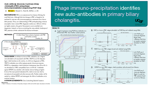
Project design: This is a case-control study of patients with primary biliary cholangitis (PBC) recruited from the UCSF liver transplant and general hepatology clinics as part of the Functional Assessment in Liver Transplantation (FrAILT) study and Prospective Observational Study to Understand Liver Disease (POSULD) cohort. Serum from 28 PBC patients and 94 healthy patients were collected and analyzed via phage immunoprecipitation sequencing (PhIP-seq). PhIP-seq allows for the quantification of auto-antibodies in patient serum in an unbiased manner. The assay was completed following methods developed by the DeRisi lab. Briefly, the assay utilizes a custom-designed phage library consisting of 49 amino acid peptides spanning the entire human proteome with 25 amino acid overlap presented on the phage capsid surface. This phage library is incubated with patient serum and the antibody-phage complexes are then immunoprecipitated. The phage are sequenced and further analyzed to map each human peptidome hit back to its human protein of origin. Analysis of sequencing reads was performed via python bioinformatics programming scripts developed in the DeRisi lab. Validation of hits was performed utilizing HiBit, whereby peptide sequences identified were synthesized with the addition of a tag (HiBit). When HiBit binds to another subunit (LgBit), the HiBit-LgBit complex contains luciferase activity which can be quantified upon addition of its substrate. These synthetic tagged peptides were incubated with patient serum and the antibody-peptide complexes were pulled down. LgBit was added with substrate and luminescence was measured.
Results: The cohort included 28 PBC patients and 94 healthy patients. 85% of PBC patients were female while 58.5% of healthy patients were female. Mean age was 61 for PBC patients and 42 for healthy controls. Racial data for the healthy cohort was incomplete as 36 patients had missing racial data. Regarding ethnicity, 57.1% of PBC and 57.4% of healthy patients identified as Hispanic. Within PBC patients, 20 patients were AMA positive and 19 patients had advanced liver fibrosis, defined as stage 3 and above. Logistic regression analysis of PhIP-seq antibodies can differentiate PBC patients from healthy controls with area under the curve (AUC) of 0.9. The highest coefficient in the logistic regression correspond to a known PBC antigen, the E2 subunit of the branched-chain alpha keto acid dehydrogenase complex (or DBT). As defined as a z-score above 3, 10 PBC patients were positive for DBT, while only 1 healthy patient was positive. Presence of auto-antibodies against this DBT peptide was validated using HiBit as 5 out of 8 PBC patients tested positive while 0 out of 5 healthy patients were positive. The second highest logistic regression coefficient corresponded to thyroid hormone receptor interactor 12 (TRIP12). Based on z-scores, the number of positive patients for TRIP 12 was 6 PBC patients and 2 healthy ones. Upon testing serum from 12 PBC patients and 10 healthy controls via HiBit, antibodies against TRIP12 were further validated as the 6 previously identified positive patients were found to have antibodies against TRIP12. Antibodies to TRIP12 were not found in other patients via HiBit. The last hit interrogated is dipeptidase 2 (DPEP2). Based on z-scores, 6 PBC patients were positive while 4 healthy patients were positive resulting in sensitivity of 21.4% and specificity of 95.7%. Out of the 6 positive PBC patients, 5 patients had advanced liver fibrosis. Unfortunately, we were unable to validate the presence of autoantibodies to DPEP2 via HiBit, which may be related to HiBit peptide folding. Discussion: Using PhIP-seq, we were able to identify known and new antibodies associated with PBC. Amongst the known ones, we detected antibodies against DBT. However, we were unable to detect antibodies against PDC-E2, the main antigen in PBC. This may be related to limitations of PhIP-seq as it can only identify antibodies against linear epitopes or those with no-post translational modifications. In fact, the primary antibody epitope of PDC-E2 is known to be posttranslationally modified via lipoylation.1 As part of the new antibodies detected, TRIP12 was identified by PhIP-seq and validated via HiBit. TRIP12 is an E3 ubiquitin ligase involved in DNA damage repair, which localizes to the nucleus. It remains unclear how antibodies against a nuclear antigens, such as TRIP12, may participate in PBC pathophysiology. As anti-nuclear antibodies are diagnostic in PBC, TRIP12 antibodies may reflect further epitope spread of the immune response in PBC. We were drawn to DPEP2 as it was identified by GWAS as a PBC susceptibility locus.2 DPEP2 is a membrane-bound dipeptidase involved in hydrolyzing leukotrienes from D4 to E4. Of note, leukotrienes E4 have decreased affinity for their receptor as compared to D4. Antibodies against DPEP2 may hinder its catalytic activity, thereby increasing the amount of a more potent leukotriene. Leukotrienes are mainly involved in eosinophil recruitment and eosinophilic infiltration has been found in ~29% of PBC liver biopsies. Additionally, anti-leukotriene therapy has been used in PBC with some mild biochemical improvements.3 Unfortunately, we were unable to validate via HiBit, possibly due to differential folding between the phage peptide and Hibit peptide. More experiments utilizing whole protein will be required for validation.
References: 1. Mao, T. K., Davis, P. A., Odin, J. A., Coppel, R. L. & Gershwin, M. E. Sidechain Biology and the Immunogenicity of PDCE2, the Major Autoantigen of Primary Biliary Cirrhosis. Hepatology 40, 1241–1248 (2004).
2. Hitomi, Y. & Nakamura, M. The Genetics of Primary Biliary Cholangitis: A GWAS and Post-GWAS Update. Genes (Basel) 14, 405 (2023).
3. Miyaguchi, S., Oda, M., Saito, H. & Ishii, H. Novel therapeutic approach to primary biliary cirrhosis patients: anti-eosinophil strategy. Hepatogastroenterology 45, 1457–1461 (1998).