145 posters, 12 topics, 25 sessions, 145 authors
ePostersLive by SciGen Technologies S.A. All rights reserved.
UCSF 2023 Inquiry Symposium
10 May | University of San Francisco

P060
A More Posterior Tibial Tubercle (Decreased Sagittal Tibial Tubercle-Trochlear Groove Distance) Is Significantly Associated With Patellofemoral Joint Degenerative Cartilage Change: A Deep Learning Analysis
Poster Presenter
Authors
Part of Topic
Clinical & Translational Sciences
Introduction
KneIntroduction
Knee osteoarthritis (OA) is a heterogenous disease process that may arise from chondral
lesions. One of the most commonly affected compartments in knee OA is the patellofemoral
joint (PFJ). Management is often non-operative, though sometimes surgery is necessary to
realign the PFJ and offload the articular surfaces. Understanding anomalies in geometric
morphology contributing to PFJ cartilage loss may provide better understanding of the natural
history of PFJ OA.
Hypothesis
It was hypothesized that an increase in anteroposterior and mediolateral distance between the
tibial tubercle and trochlear groove is associated with PFJ OA, decreased patellar cartilage
thickness, and increased patellar cartilage T2 relaxation time.
Aims
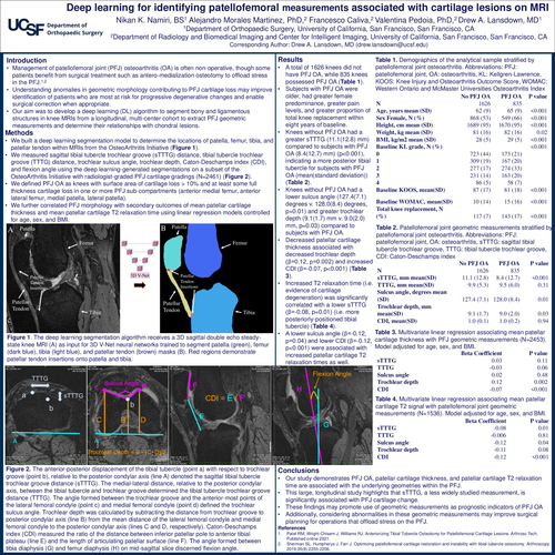
To perform PFJ geometric measurements on knee magnetic resonance imaging scans and
determine their relations with chondral lesions in a multicenter cohort using deep learning.
Methods
The sagittal tibial tubercle–trochlear groove (sTTTG) distance, tibial tubercle–trochlear groove
distance, trochlear sulcus angle, trochlear depth, Caton-Deschamps Index (CDI), and flexion
angle were measured by use of deep learning–generated segmentations on a subset of the
Osteoarthritis Initiative study with radiologist-graded PFJ cartilage grades (n = 2,461). Kruskal-
Wallis H tests were performed to compare differences in PFJ morphology between subjects
without PFJ OA and those with PFJ OA. PFJ morphology was correlated with secondary
outcomes of mean patellar cartilage thickness and mean patellar cartilage T2 relaxation time
using linear regression models controlling for age, sex, and body mass index.
Results
A total of 1,626 knees did not have PFJ OA, whereas 835 knees had PFJ OA. Knees without
PFJ OA had an increased (anterior) sTTTG distance (mean ± standard deviation, 11.1 ± 12.8
mm) compared with knees with PFJ OA (8.4 ± 12.7 mm) (P < .001), indicating a more posterior
tibial tubercle in subjects with PFJ OA. Knees without PFJ OA had a decreased sulcus angle
(127.4° ± 7.1° vs 128.0° ± 8.4°, P = .01) and increased trochlear depth (9.1 ± 1.7 mm vs 9.0 ±
2.0 mm, P = .03) compared with knees with PFJ OA. Decreased patellar cartilage thickness was
associated with decreased trochlear depth (β = 0.12, P = .002) and increased CDI (β = –
0.07, P < .001). Increased patellar cartilage T2 relaxation time was correlated with decreased
sTTTG distance (β = –0.08, P = .01), decreased sulcus angle (β = –0.12, P = .04), and
decreased CDI (β = –0.12, P < .001).
Discussion
Our study demonstrates PFJ OA, patellar cartilage thickness, and patellar cartilage T2
relaxation time are associated with the underlying geometries within the PFJ joint. TTTG, sulcus
angle, trochlear depth, and CDI have been previously investigated for associations with
chondral defects. We expand on this body of literature with a large, longitudinal sample while
also demonstrating sTTTG, a less widely studied measurement, significantly contributes to PFJ
cartilage change. These findings may promote the use of geometric measurements as
prognostic indicators of PFJ OA. Additionally, considering abnormalities in these geometric
measurements may improve surgical planning for operations that offload stress on the PFJ.
Our study contains limitations, beginning with definitions of our study outcomes. Subjects were
classified as having PFJ OA, the primary outcome, if they possessed cartilage loss in either the
patella or trochlea. However, the study’s secondary outcomes (i.e. patellar cartilage thickness
and patellar cartilage T2 relaxation time) were only measured on the patella, not trochlea. The
inability of this study to include trochlea during measurement of secondary outcomes may
explain why some PFJ measurements like CDI were significantly associated with secondary
outcomes (i.e. patella cartilage thickness and patellar cartilage T2 relaxation time) but not the
primary outcome, PFJ OA, which is a measure of both patella and trochlea. Moreover, even
though differences in measurements proved statistically significant, such as the difference in
sTTTG between controls and cases with PFJ OA, they may not necessarily warrant clinical
significance.