145 posters, 12 topics, 25 sessions, 145 authors
ePostersLive by SciGen Technologies S.A. All rights reserved.
UCSF 2023 Inquiry Symposium
10 May | University of San Francisco

P012
Modeling Risk of Progression to Lung-RADS 4 from a Benign Lung Cancer Screening CT using Imaging and Clinical Data
Poster Presenter
Authors
Part of Topic
Clinical & Translational Sciences
Introduction:
Annual screening for asymptomatic lung cancer with low-dose chest CT scans (LDCTs) in
eligible people is an evidence-based practice for reducing lung cancer mortality. LDCTs are
often scored using the Lung-RADS guidelines, which recommend interval of imaging and clinical
workup. The interval of next screening is standardized to one year for patients with benign
LDCTs (Lung-RADS ≤ 2), representing ~90% of all screenings. However, at the next annual
screen, some such patients may continue to have low risk LDCTs while others may present with
findings highly suggestive of cancer. Previously developed models for more accurate future lung
cancer risk estimation do not provide tailored predictions based on Lung-RADS stratifications
and do not incorporate the wealth of information found in raw imaging.
Hypothesis or question:
We hypothesize that an AI model trained on demographic, clinical, and baseline imaging
characteristics can estimate the risk of benign LDCTs at baseline screen progressing to LungRADS 4 at follow-up screen, a prediction that can be used to guide individualized risk
assessment and screening interval.
Aims:
• Build statistical models for predicting progression to Lung-RADS 4 from clinical
information and baseline imaging findings
• Build a deep learning model for predict progression to Lung-RADS 4 from full-volume CT
• Evaluate generalizability of models to UCSF lung screening datasets
Project Design/Methods:
This is a retrospective modeling study that utilized clinical characteristics and imaging from the
National Lung Screening Trial (NLST) dataset as a training data. A proprietary AI-based system
from United Imaging Intelligence was used to calculate the Lung-RADS score for each scan.
Benign LDCTs (Lung-RADS ≤ 2) were selected. For the clinical and demographics data,
recursive feature selection was conducted to select the four most predictive features. Then,
statistical and deep learning models were developed, trained, and compared based on the
AUC-ROC score. Finally, we evaluated generalization to clinical and imaging data from benign
UCSF’s lung cancer screenings.
Results:
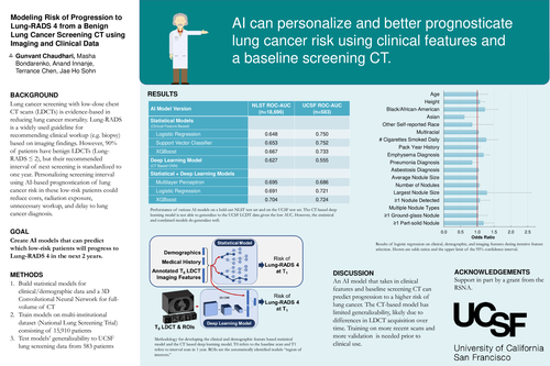
From the NLST dataset, a subset 18,696 baseline LDCT screenings were found to have LungRADS ≤ 2, with 1,837 progressing to Lung-RADS 4 (9.8%). Similar benign Lung-RADS score
studies from UCSF totaled 583 studies, with 82 (14%) progressing to Lung-RADS 4 in two years.
From a logistic regression predicting Lung-RADS progression, features that were significantly
associated were height, emphysema diagnosis, number of nodules, and pack-year history. From
these features, the best performing statistical model for predicting progression had an AUC of
0.627 on the NLST data and 0.750 on the UCSF data. The deep learning-based model trained on
full volume CTs had an AUC of 0.627 and 0.555 on the NLST and UCSF data, respectively. A
combined model with clinical features and full-volume CT had a maximal AUC of 0.704 and
0.724 on the NLST and UCSF data, respectively.
Discussion/Conclusion:
This study shows that an AI based model that takes in clinical features and full-volume CT scan
can predict progression to Lung-RADS 4 with a maximal AUC of 0.704, and that this bestperforming model generalizes to UCSF data with an AUC of 0.724. One limitation is that the
deep learning model based on the full CT volume had a significant drop in performance when
tested on the UCSF data. This may be due to differences in low-dose CT acquisition over time
as the NLST dataset is from the early 2000s, while the UCSF dataset includes data up to 2022.
More investigation on improving generalizability of this system will be necessary to create an
accurate model that clinicians can use to personalize their patients’ interval of lung cancer
screening.