145 posters, 12 topics, 25 sessions, 145 authors
ePostersLive by SciGen Technologies S.A. All rights reserved.
UCSF 2023 Inquiry Symposium
10 May | University of San Francisco

P053
Detection of incidental malignancy on Prostate Specific Membrane Antigen (PSMA) PET/CT imaging: a review of 2,054 sequential studies at an academic medical center
Poster Presenter
Authors
Part of Topic
Clinical & Translational Sciences
Introduction:
Prostate specific membrane antigen (PSMA) is a transmembrane glycoprotein that has been shown to
be highly expressed in prostate tissue, with particularly high uptake in primary prostate cancer and
lymph node metastases [1]. However, PSMA PET uptake has been described in many case reports of
non-prostate cancer lesions, including various non-prostate malignancies and benign processes [2]–[11].
There have been limited prior studies that systematically examined a series of PSMA PET/CT studies for
PSMA-ligand avidity corresponding to non-prostate cancer incidental findings [12], [13]. This raises the
question of which PSMA-ligand avid incidental lesions are malignant and would thus require follow-up
that would affect clinical management.
Hypothesis or question (if applicable):
From review of reports with similar radiotracers, PSMA-ligand avidity is expected to be the highest in
prostate tissue, particularly primary prostate cancer and lymph node metastases. However, incidental
lesions with PSMA-ligand avidity are expected to be found in as many as 10% of cases, with about an
even distribution of confirmed prostate metastatic, secondary metastatic, and benign lesions on those
with a follow-up biopsy [12], [13].
Aims:
This study aims to catalog confirmed the types of non-prostate malignancies detected by PSMA PET/CT.
The aim was also to identify imaging features that may distinguish secondary malignancies from
prostate metastasis and benign lesions, or benign lesions from any malignant lesions.
Project Design/Methods:
A retrospective review of the UCSF mPower database of all UCSF 68 Ga-PSMA-11 (PSMA) PET/CT studies
completed from December 2015 - April 2022 was performed. This yielded a total of 2,054 imaging
reports (1873 unique patients). Reports that noted incidental PSMA avidity not definitively related to
previously known prostate cancer distribution or physiologic uptake were included, resulting in a set of
255 imaging reports (245 unique patients). The final cohort was composed of patients who additionally
had pathology reports from completed biopsies available within APeX, resulting in the study population
of 50 studies from 49 patients with a history of prostate cancer, where one patient had workup for two
distinct lesions from separate PSMA PET/CT studies.
All images were retrospectively reviewed in separate sessions by a junior and a senior radiologist, who
were unaware of the pathological diagnosis of the lesions. Lesions were judged quantitatively by several
SUV metrics and characterized qualitatively, measured separately by both readers. Discrepancies in
lesion determination were resolved by consensus. Categorical parameters, such as lesion location and
the indication for the PSMA PET/CT study, were compared by multinomial test and binomial test.
Continuous parameters, such as SUVmax and size of the lesions, were compared by Kruskal-Wallis test
and Wilcox test. P values of less than 0.05 were considered statistically significant.
Results/Product:
Of the imaging records reviewed, 50 PSMA PET/CT studies of 49 patients with a history of prostate
cancer were found to have incidental findings that resulted in diagnostic biopsy of 50 distinct lesions. 11
of these PSMA PET/CT studies were for a new diagnosis of prostate cancer, and 39 were for biochemical
recurrence. The median age at the time of imaging was 72 years old (range: 58-92, SD: 7.32) and the
median PSA at the time of imaging was 3.555 (range: 0.06 – 103, SD: 18.63). 10 patients (20%) had a
diagnosis of a non-prostate malignancy prior to PSMA PET/CT. 4 patients (8%) had a prior PSMA PET/CT
study.
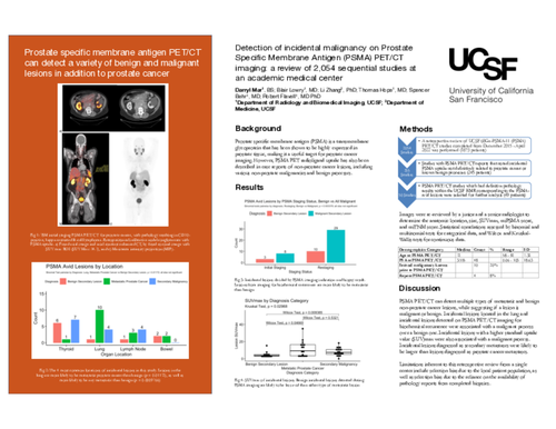
A total of 20 secondary malignancies, 17 prostate cancer metastases, and 13 benign lesions were
revealed. PSMA-ligand avid incidental lesions were found in a variety of organ locations. These were the
lung (15), thyroid (14), lymph node (8), bowel (4), liver (2), and 1 each in tissue of the muscle, the renal
system, the bladder, the bone, the neuroforamina, the tongue, and a distinct mass. Lesions in the lung
were more likely to be metastatic than benign (p = 0.0009766). All other comparisons amongst lesions in
the same organ were insignificant. Incidental PSMA-ligand avid lesions found on PSMA PET/CT studies
performed for new prostate cancer were more likely to be a secondary malignancy than a metastatic
prostate lesion (p = 0.007812). Incidental PSMA-ligand avid lesions found on PSMA PET/CT studies
performed for biochemical recurrence were more likely to be malignant secondary lesions than benign
(p = 0.003378). Benign secondary lesions were less PSMA-ligand avid than both metastatic prostate
lesions (p = 0.04665) and secondary malignancies (p = 0.009385). Benign lesions also exhibited lower
miPSMA scores (p = 0.01742) than metastatic prostate lesions. Secondary malignancies were larger than
metastatic prostate cancer lesions when measured by their longest dimension (p = 0.0007943). PSA and
age at the time of imaging were not associated with the diagnosis of the incidental lesion.
Discussion/Conclusion:
Our study demonstrates that PSMA-ligand avid incidental lesions can be found in a variety of tissues,
primarily the lung, thyroid, and lymph nodes. Several factors may influence the probability that an
identified lesion is a secondary malignancy, a prostate malignancy, or benign. These included the
locations of the lesions and whether the PSMA PET/CT study was performed for new or biochemically
recurrent prostate cancer. The finding that PSMA-ligand avid benign secondary lesions are significantly
less avid than malignant lesions is consistent with the limited other studies that have examined
incidental lesions from PSMA PET imaging [12], [13]. This study has the potential to guide clinical
decision making on further diagnostic workup in patients who have incidental PSMA PET/CT findings.
The study had several limitations. First, the study used patient data from a single center, introducing
selection bias based on the patient population of the center. There may be further selection bias due to
the reliance on the original reading radiologists of the studies to consistently identify suspicious lesions
inconsistent with known patterns of metastatic prostate or benign PSMA-ligand uptake. Another
important factor is the reliance on the availability of diagnostic biopsies for patient follow-up. This may
reflect a selection bias based on the performance of biopsies for patients for whom the index of
suspicion for a secondary malignancy is high, or for whom the potential to alter clinical management is
greatest.