145 posters, 12 topics, 25 sessions, 145 authors
ePostersLive by SciGen Technologies S.A. All rights reserved.
UCSF 2023 Inquiry Symposium
10 May | University of San Francisco

P042
Comparison of Retinal Degeneration in GUCY2D and CRX-related Autosomal Dominant Cone-Rod Dystrophy
Poster Presenter
Authors
Part of Topic
Clinical & Translational Sciences
Purpose To compare cone structure and function in patients with autosomal dominant-cone
rod dystrophy (AD-CRD) due to genetic mutations in retinal guanylate cyclase (GUCY2D),
encoding a protein that modulates calcium metabolism, and in the homeodomain transcription
factor (CRX) that regulates photoreceptor differentiation.
Methods Refractive error, best corrected visual acuity, spectral domain optical coherence
tomography (SD-OCT), color and short-wavelength autofluorescence fundus photos were
obtained in all subjects. Confocal and split-detector adaptive optics scanning laser
ophthalmoscopy (AOSLO) images were acquired in one patient each with GUCY2D and CRX with
stable fixation. Cone spacing was calculated using a density recovery profile method and Z-
scores to report standard deviation (std dev) from the normal mean at the eccentricity of the
measured cones; < 2 was within normal limits. Differences between groups were assessed by
Student’s t-test (GraphPad PRISM-9 software).
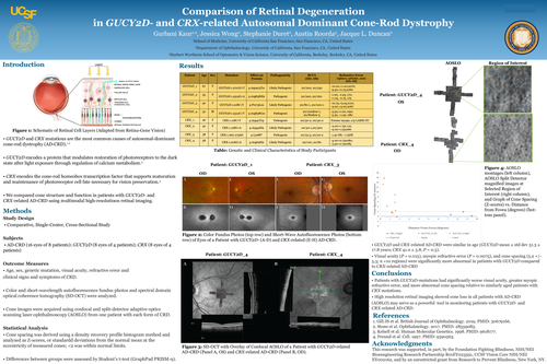
Results Our cross-sectional study included 14 eyes of 7 ADCRD patients (3 with GUCY2D and 4
with CRX mutations; the groups were not different in age (GUCY2D mean±std dev 51.3 ± 17.8
years, and CRX 41.0 ± 3.8, P = 0.3). Though both groups displayed reduced visual acuity and
central scotomas, CRX patients had better visual acuity (P = 0.013) and less myopic refractive
error (P = 0.0075) than GUCY2D patients. Fundus autofluorescence revealed retinal pigment
epithelial mottling in early stages and bull’s eye maculopathy in advanced disease in both
groups. SD-OCT revealed outer retinal layer thickness loss with foveal atrophy in all but one
GUCY2D patient and ellipsoid zone irregularity in CRX patients. AOSLO images showed no
evidence of cone structure at the anatomical fovea for either CRX- or GUCY2D-related AD-CRD.
In the patient with CRX-related AD-CRD, cone spacing Z scores were within normal limits (mean
± std dev = 0.76 ± 0.70; n=3 regions), while in the eyes with GUCY2D-related AD-CRD, Z-scores
were abnormal (5.2 ± 5.3; n=10 regions).
Conclusions AD-CRD may differ in disease severity as evidenced by worse visual acuity, greater
myopic refractive error and more abnormal cone spacing in GUCY2D patients relative to
similarly aged CRX patients. High resolution retinal imaging showed cone loss in all imaged
patients and may be useful for diagnosing and monitoring disease severity in patients
with GUCY2D and CRX-related AD-CRD.