145 posters, 12 topics, 25 sessions, 145 authors
ePostersLive by SciGen Technologies S.A. All rights reserved.
UCSF 2023 Inquiry Symposium
10 May | University of San Francisco

P018
Lung Nodule Prediction Tool for use in Areas Endemic to Coccidioidomycosis
Poster Presenter
Authors
Part of Topic
Clinical & Translational Sciences
Title
Lung Nodule Prediction Tool for use in Areas Endemic to Coccidioidomycosis
Introduction
Lung nodules are common. They may represent lung cancer or may be due to endemic fungi including
coccidioidomycosis (cocci). Currently validated lung nodule risk calculators inappropriately classify 84-
94% of cocci nodules as intermediate-to-high risk for lung cancer. This leads to additional workup
associated with unnecessary costs and complications.
Aim
Develop a prediction tool to differentiate lung nodules due to lung cancer from those due to cocci in
areas endemic to cocci.
Project Design/Methods
Study design: We identified patients seen in the UCSF Fresno multidisciplinary lung nodule program who
were diagnosed with stage I-IIIB lung cancer or cocci between 01/01/2011 and 10/31/2022. Patients
were identified using ICD codes. We excluded patients diagnosed with stage IV lung cancer or without
an official radiology report in our electronic medical record (EMR). Patients diagnosed with cocci were
categorized as proven (biopsy, culture, or PCR positive), probable (radiographic and clinical stability in
the setting of necrotizing granuloma on biopsy or positive cocci serology), or possible (large nodule
without an alternative diagnosis with clinical and/or radiographic stability). We conducted chart reviews
to collect candidate predictor variables including clinical characteristics, radiographic findings, and
serologic test results. Radiographic information was collected from official radiology reports in our EMR.
This study was approved by our institutional review board (IRB# 2022019).
Statistical analysis: Descriptive statistics described the sample characteristics of patients with lung
cancer and cocci. A random ⅔ sample of the data set was used as the training data set. The remaining ⅓
served as the validation data set. Bivariate analyses were performed on the training data set using
simple logistic regression. Predictors with P<0.05 based on the bivariate analysis were included in the
development of the multivariable model. Stepwise, backward multivariable logistic regression models
were performed, with the final model containing plausible predictors with a P-value <0.05. Goodness of
model fit was evaluated using a Hosmer-Lemeshow test. The predictive performance of the integer
clinical prediction score in predicting lung cancer vs. cocci was assessed by using the area under the
Receiver Operating Characteristic (ROC) curve (AUC) in the validation cohort. Sensitivity and specificity
were evaluated at each cut-point with 95% confidence interval (CI) estimated. Prediction models derived
using the training data set were validated by means of an assessment of discrimination accuracy in the
validation data set. Statistical analyses were performed using Stata (StataCorp. 2021. Stata Statistical
Software: Release 17. College Station, TX: StataCorp LLC).
Results
We identified 806 patients who met our inclusion criteria - 566 patients diagnosed with stage I-IIIB lung
cancer and 240 patients diagnosed with cocci. We excluded 319 patients with stage IV lung cancer and
23 patients without a radiology report in our EMR (8 cancer, 15 cocci) because their imaging was done
outside our clinic. Of the patients diagnosed with cocci, 125 (52.1%) had proven cocci, 111 (46.3%)
probable, and 4 (1.7%) possible. 150/240 (62.5%) patients with cocci underwent CT-guided or
transbronchial biopsy and 70/240 (29.2%) underwent BAL. 164/240 (68.3%) underwent one or both
invasive procedures. A random ⅔ sample (N=532) was used as the training data set and the remaining ⅓
(N=274) served as the validation data set.
Bivariate analyses of clinical, radiographic, and serologic candidate predictors using the training data set
were performed. Eight clinical characteristics (age, sex, pack years, personal history of malignancy,
family history of lung cancer in a 1st degree relative, clinical history of chronic lung disease, work in
agriculture, and exposure to asbestos), 6 radiographic findings (nodule location, density, border
characteristics, cavitation, satellite nodules, and radiographic evidence of chronic lung disease), and
both serologic tests (immunodiffusion (ID) and complement fixation (CF)) were independent predictors,
with P<0.05. These predictors – except for ID and CF, which contained excess missing data and were
excluded to prevent selection bias – were used in a stepwise logistic regression analysis to derive a
multivariable prediction model. ROC curves were developed for the training and validation data sets.
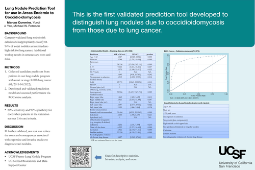
The results of the ROC curve analyses showed good overall discrimination accuracy for both the training
(AUC = 0.91; 95% CI, 0.88-0.93) and validation data sets (AUC = 0.90; 95% CI, 0.86-0.94). The optimum
cutoff point identified by Youden’s test was 6. When patients in the validation data set met ≥ 6 of the
following criteria – age < 60, male sex, < 50 pack years, no exposure to asbestos, no ground glass
component(s), right middle or left upper lobe, no spiculated/lobulated/irregular borders, cavitation,
satellite nodules, no radiographic evidence of chronic lung disease – there was a sensitivity of 0.80 (95%
CI, 0.72-0.89) and specificity of 0.90 (95% CI, 0.85-0.94) for diagnosing the nodule as due to cocci.
Discussion
Our prediction tool successfully predicted 80% of cocci nodules while only falsely labeling 10% of lung
cancer nodules as cocci. The 20% of cocci nodules that the tool failed to detect represents a 71%
reduction from the 68.3% of patients with cocci in our cohort who underwent an invasive procedure
(biopsy and/or BAL). After a nodule has been deemed intermediate-to-high risk for lung cancer using
one of the previously validated calculators, our tool can be used in areas endemic to cocci to determine
whether additional invasive diagnostic workup is necessary.
Our study has a few limitations. Information available in the official radiology report limited our
collection of radiographic data. About half of radiology reports did not describe the border characteristic
of the nodule. The borders in these cases may have been difficult to determine or not concerning to the
radiologist. If the imaging were reviewed, border characteristics may have been determined. However,
we wanted to be consistent in our data collection process and relied only on the information available in
the radiology report. As mentioned previously, ID and CF contained excess missing data and were
excluded from multivariable model development to prevent selection bias. We plan to further
investigate integrating serologic studies into our prediction tool and hope to determine which patients
would benefit most from serologic workup. Another limitation of our study is the single-center design.
Our results may not generalize to other areas endemic to cocci. However, our study may encourage
other institutions to evaluate our tool or develop similar prediction tools. It may also serve as a
framework for the development of models in geographic areas endemic to other dimorphic fungi, such
as blastomycosis and histoplasmosis.
This is the first validated prediction tool developed to distinguish lung nodules due to cocci from those
due to lung cancer. It utilizes standard clinical and radiographic data, and, if further validated, can
significantly reduce the costs and complications associated with expensive and invasive studies to
diagnose cocci nodules.