334 posters, 125 sessions, 831 authors, 236 institutions
ePostersLive by SciGen Technologies S.A. All rights reserved.
78th Annual PostGraduate Assembly in Anesthesiology (PGA78) 2024
December 6 - 9, 2024 | New York City

3092
External Ultrasound Guided Trigger Point Injection for Female Patient with Hypertonic Pelvic Floor Dysfunction
Poster Presenter
Authors
Affiliations
Part of Topic
Ultrasound Guided External Trigger Point Injections for Myofascial Pelvic Pain in a Female Patient
A widely-used clinical definition for chronic pelvic pain is noncyclical pelvic pain that lasts for at least six months.1 As is with most chronic pain conditions, there are multifactorial contributors to the pain experience. Although a diagnosis of pelvic pain may be made, there are often a number of individual contributors such as myofascial components and central sensitization. High-tone pelvic floor disorder (HTPFD), also known as myofascial pelvic pain syndrome, is one of the most common contributing cause of chronic pelvic pain in women.2 HTPFD is characterized by areas of hypertonicity, or abnormally increased resting muscle tone or tightness, of the pelvic floor muscles.3-5
Myofascial trigger points, which are localized areas of tenderness within a taut portion of skeletal muscle, may occur in hypertonic muscles.2,6-8 Trigger points may be a result of muscle injury, which may include a perceptible trauma or repeated microtrauma, or may occur without a clear inciting incident.7-8 Trigger points are best identified upon physical examination because they feel taut when palpated perpendicular to the fiber direction and are painful when compressed.2,7
In 2024, Torosis et al. published a consensus-based treatment algorithm for HTPFD. Providers agreed that first-line treatment for HTPFD should be pelvic floor physical therapy (PFPT), second-line treatment includes trigger point injections (TPIs), vaginal muscle relaxants, and cognitive behavioral therapy, third-line treatment consists of botulinum toxin injections, and sacral neuromodulation is a fourth-line therapy. In this case we focus on TPIs for treatment of HTPFD, which are best used in conjunction with other therapies.
TPIs are used to relieve myofascial pain via injection of trigger points.8 Trigger points may be injected with a needle alone (dry needling), a local anesthetic with or without adding a corticosteroid, or botulinum toxin.8-9 At this time, there is no definitive evidence suggesting superiority of one injectate over the others.8,10 TPIs may also be done with or without the guidance of imaging, such as fluoroscopy and ultrasound.8 Studies have suggested that there are benefits to using ultrasound to guide injections including increased accuracy and avoidance of complications from blind injections.8,11,12 TPIs targeting pelvic floor muscles can be external (pelvic) or internal (vaginal / transvaginal), and different approaches may be employed depending on the muscle groups being targeted. A transvaginal approach can be used in female patients to access deeper muscles such as the obturator internus, obturator externus, and levator ani, while an external approach can be used to access superficial muscles including the gluteus, bulbospongiosus, and transverse perineal muscles.
There has been no retrospective or prospective study that has comprehensively evaluated the use of external TPIs for pelvic floor dysfunction. Here, we present a case where ultrasound-guided external TPIs were successfully used to relieve myofascial pelvic pain due to HTPFD in a female patient.
A 30-year-old woman presented to our clinic with myofascial pelvic pain due to HTPFD. The patient had a 15-year history of pelvic pain of multiple etiologies including vaginismus and vulvodynia (s/p valvulectomy). The patient had a fall 20 years ago on her tailbone and had tailbone pain at the time of the visit. The pelvic pain was sharp in the perineal area and would shoot forward. The patient’s pain was worse with intercourse and menstruation. The pain was made better by consistent yoga practice and ibuprofen. The patient tried PFPT and biofeedback with no improvement. The patient also tried diazepam suppositories which provided relief. She received a botulinum toxin injection two years prior to presentation to our clinic which provided some relief. She had a pudendal nerve block one year prior to presentation to our clinic, which provided 40% relief for 4-5 months.
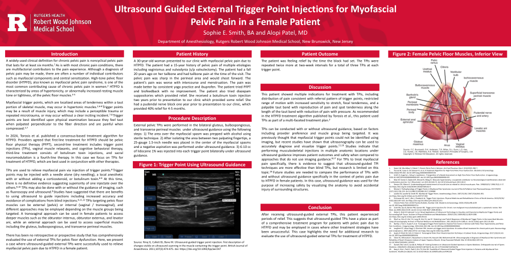
External pelvic TPIs were performed in the bilateral gluteus, bulbospongiosus, and transverse perineal muscles under ultrasound guidance using the following steps: 1) The area over the myofascial spasm was prepped with alcohol using sterile technique. 2) After isolating the area between two palpating fingertips, a 25-gauge 1.5-inch needle was placed in the center of the myofascial spasms and a negative aspiration was performed under ultrasound guidance. 3) 6-10 cc of lidocaine 1% was injected into each trigger point bilaterally under ultrasound guidance.
The patient was feeling relief by the time the block had set. The TPIs were repeated twice more at two-week intervals for a total of three TPIs at each trigger point.
This patient showed multiple indications for treatment with TPIs, including distribution of pain consistent with referral pattern of trigger points, restricted range of motion with increased sensitivity to stretch, focal tenderness, and a palpable taut band with reproduction of pain and spot tenderness along the length of the taut band with reduction of pain with pressure. As recommended in the HTPFD treatment algorithm published by Torosis et al., this patient used TPIs as part of a multi-faceted treatment plan.2
TPIs can be conducted with or without ultrasound guidance, based on factors including provider preference and muscle group being targeted. It was previously thought that myofascial trigger points could not be identified using imaging, but recent studies have shown that ultrasonography can be used to accurately diagnose and visualize trigger points.11,16 Studies indicate that performing musculoskeletal injections in multiple anatomic locations under ultrasound guidance improves patient outcomes and safety when compared to approaches that do not use imaging guidance.8,17 For TPIs to treat myofascial pain specifically, there is evidence to suggest that ultrasound-guided TPI techniques are more effective than blind TPIs, but research is limited on this topic.18 Future studies are needed to compare the performance of TPIs with and without ultrasound guidance specifically in the context of pelvic pain due to HTPFD in female patients. In this case, ultrasound guidance was used for the purpose of increasing safety by visualizing the anatomy to avoid accidental injury of surrounding structures.
After receiving ultrasound-guided external TPIs, this patient experienced periods of relief. This suggests that ultrasound-guided TPIs have a place as part of a comprehensive treatment program for patients with pelvic pain due to HTPFD and may be employed in cases where other treatment strategies have been unsuccessful. This case highlights the need for additional research to evaluate the use of ultrasound-guided external TPIs for treatment of HTPFD.