334 posters, 125 sessions, 831 authors, 236 institutions
ePostersLive by SciGen Technologies S.A. All rights reserved.
78th Annual PostGraduate Assembly in Anesthesiology (PGA78) 2024
December 6 - 9, 2024 | New York City

3079
An Unexpected Twist: A Rare Case of Lung Torsion After VATS Lobectomy
Part of Topic
Learning Objective
Intra-operative and post-operative management of lung torsion as a rare complication of Video-Assisted Thoracic Surgery (VATS).
Introduction
VATS has rapidly become the gold standard over open thoracotomy for lung cancer surgery
Anesthesia perioperative planning includes: general anesthesia, double lumen tube, and one lung ventilation, standard ASA monitoring +/- arterial line, and bronchoscopy
Multimodal analgesia including thoracic epidural or paravertebral block
Common post-operative complications include atelectasis, pneumothorax, hemothorax, pneumonia, aspiration, PE, arrhythmias, and respiratory insufficiency
Rare complications include lung torsion
Case Description
A 72-year-old man with a 50 pack-year smoking history, COPD, and biopsy-proven right upper lobe NSCLC underwent VATS right upper lobectomy with one-lung ventilation using a left-sided double-lumen tube. The surgery required multiple vein isolations and parenchymal dissections to search for carcinoma. The patient remained hemodynamically stable intraoperatively with no anesthesia complications.
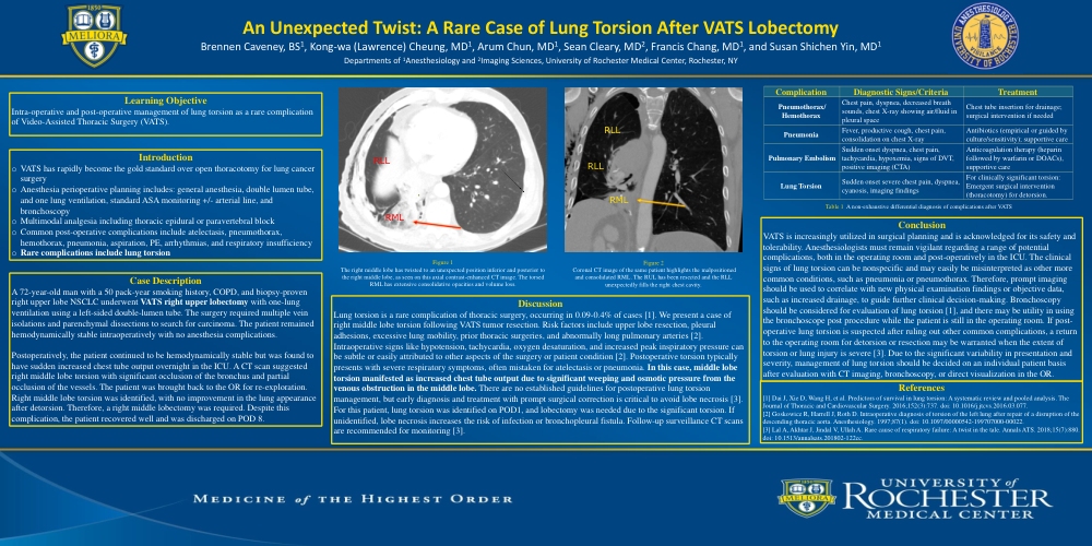
Postoperatively, the patient continued to be hemodynamically stable but was found to have sudden increased chest tube output overnight in the ICU. A CT scan suggested right middle lobe torsion with significant occlusion of the bronchus and partial occlusion of the vessels. The patient was brought back to the OR for re-exploration. Right middle lobe torsion was identified, with no improvement in the lung appearance after detorsion. Therefore, a right middle lobectomy was required. Despite this complication, the patient recovered well and was discharged on POD 8.
Discussion
Lung torsion is a rare complication of thoracic surgery, occurring in 0.09-0.4% of cases [1]. We present a case of right middle lobe torsion following VATS tumor resection. Risk factors include upper lobe resection, pleural adhesions, excessive lung mobility, prior thoracic surgeries, and abnormally long pulmonary arteries [2]. Intraoperative signs like hypotension, tachycardia, oxygen desaturation, and increased peak inspiratory pressure can be subtle or easily attributed to other aspects of the surgery or patient condition [2]. Postoperative torsion typically presents with severe respiratory symptoms, often mistaken for atelectasis or pneumonia. In this case, middle lobe torsion manifested as increased chest tube output due to significant weeping and osmotic pressure from the venous obstruction in the middle lobe. There are no established guidelines for postoperative lung torsion management, but early diagnosis and treatment with prompt surgical correction is critical to avoid lobe necrosis [3]. For this patient, lung torsion was identified on POD1, and lobectomy was needed due to the significant torsion. If unidentified, lobe necrosis increases the risk of infection or bronchopleural fistula. Follow-up surveillance CT scans are recommended for monitoring [3].
Conclusion
VATS is increasingly utilized in surgical planning and is acknowledged for its safety and tolerability. Anesthesiologists must remain vigilant regarding a range of potential complications, both in the operating room and post-operatively in the ICU. The clinical signs of lung torsion can be nonspecific and may easily be misinterpreted as other more common conditions, such as pneumonia or pneumothorax. Therefore, prompt imaging should be used to correlate with new physical examination findings or objective data, such as increased drainage, to guide further clinical decision-making. Bronchoscopy should be considered for evaluation of lung torsion [1], and there may be utility in using the bronchoscope post procedure while the patient is still in the operating room. If post-operative lung torsion is suspected after ruling out other common complications, a return to the operating room for detorsion or resection may be warranted when the extent of torsion or lung injury is severe [3]. Due to the significant variability in presentation and severity, management of lung torsion should be decided on an individual patient basis after evaluation with CT imaging, bronchoscopy, or direct visualization in the OR.
References
[1] Dai J, Xie D, Wang H, et al. Predictors of survival in lung torsion: A systematic review and pooled analysis. The Journal of Thoracic and Cardiovascular Surgery. 2016;152(3):737. doi: 10.1016/j.jtcvs.2016.03.077.
[2] Goskowicz R, Harrell J, Roth D. Intraoperative diagnosis of torsion of the left lung after repair of a disruption of the descending thoracic aorta. Anesthesiology. 1997;87(1). doi: 10.1097/00000542-199707000-00022.
[3] Lal A, Akhtar J, Jindal V, Ullah A. Rare cause of respiratory failure: A twist in the tale. Annals ATS. 2018;15(7):880. doi: 10.1513/annalsats.201802-122cc.