334 posters, 125 sessions, 831 authors, 236 institutions
ePostersLive by SciGen Technologies S.A. All rights reserved.
78th Annual PostGraduate Assembly in Anesthesiology (PGA78) 2024
December 6 - 9, 2024 | New York City

3051
Management of Severe Airway Stenosis in a 18-Year-Old Patient at a Small District Hospital: A Case Report
Poster Presenter
Part of Topic
Emergency Management of Severe Subglottic Airway Stenosis in an 18
Hospital:A
Shady Ekladious, MD1; Hamid Raoof, MD2; Andre Van Zundert, MD, PhD3; Adam Eslick, MD4 University of Queensland, Gladstone Hospital Central Queensland Health Australia
-
Year
-
Old at a Rural District
Case Report
CASE DETAILS
|
An 18-year-old female initially presented to the rural hospital emergency department with sore throat, dysphonia and headaches. She returned two days later presented via ambulance with severe stridor, progressive dyspnea, difficulty swallowing saliva, and increasing throat pain after a recent presumed laryngitis. Initial management by the paramedics included treatment for an airway emergency and possible anaphylaxis, with pre-hospital administration of 500 mcg intramuscular adrenaline, a 5 mg nebulized adrenaline, and loratadine, leading to some stridor improvement. Despite this, she remained in respiratory distress: |
|
|
Assessment |
Findings |
|
Airway (A) |
Stridor noted on coughing and deep inspiration; voice completely hoarse; unable to tolerate secretions |
|
Breathing (B) |
Tachypneic with respiratory rate in high 20s; oxygen saturation maintained |
|
Circulation (C) |
Tachycardic (130-140 bpm, likely due to sepsis/adrenaline); robust blood pressure |
|
Disability (D) |
Fully alert (GCS 15); no focal neurological deficits |
|
Exposure (E) |
Non-blanching rash on bilateral calves; reddish rash on right chest wall; swollen lower lip |
ABOUT THE AUTHOR
Dr. Shady Ekladious is an Australian Anaesthesiology registrar with training in NICU, ICU, and anaesthetics, having trained across various city and regional areas of Australia. He is committed to equitable healthcare access and serves as a Conjoint Associate Lecturer and Clinical Tutor at several universities. Passionate about advancing medical education, Dr. Ekladious has a special interest in difficult airways, research, and medical education.
IMAGING
MANAGEMENT DISCUSSION
Initial Management:
The anaesthetic team administered dexamethasone and adrenaline nebulizers, which helped stabilize her symptoms. Oxygen saturation improved to 98%, but the team remained concerned about her airway given the 3 mm stenosis and lack of onsite ENT support.
A multidisciplinary team discussion took place involving the rural anaesthetic team, retrieval services, and an ENT surgeon from the nearest tertiary centre. A plan was established to intubate the patient under awake fibreoptic guidance, with preparations for an emergency front-of-neck access if required. The ENT surgeon arrived with the retrieval team via helicopter, ready to perform an emergency tracheostomy if necessary.
Intervention and Transfer:
The patient was successfully intubated with an awake fibreoptic approach, allowing safe transport to a tertiary ICU. The ENT surgeon’s preparedness for an emergency tracheostomy contributed to the patient’s safety during this high-risk procedure.
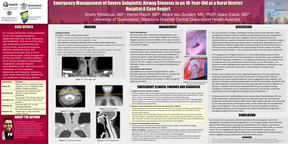
Imaging Findings:
An urgent CT neck with contrast showed
- There is significant oedematous changes of the glottis and subglottis bilaterally,
causing severe stenosis of the subglottic airway.
- The maximum transverse diameter of the airway is 3mm at this level
- There are multiple enlarged generalised cervical, bilateral axillary and hilar lymph
nodes suggestive reactive lymphadenopathy.
- Patchy airspace opacities in the left upper lobe and subtle opacity in the right
upper lobe suggesting infective or inflammatory pathology.
- Short segment of thrombosis in right sigmoid dural sinus.
Figure 1. CT neck sagittal view .
Figure 6. Inflamed airway visualized
Figure 7. ETT inserted successfully in inflamed airway via fiberoptic.
SUBSEQUENT CLINICAL FINDINGS AND DIAGNOSIS
• This case presents a complex, multifaceted medical scenario that intersects airway management, infectious disease, autoimmune pathology, and vascular complications. The initial presentation with severe subglottic stenosis, marked by stridor and respiratory distress, required immediate intervention and diagnostic clarity to manage a rapidly deteriorating airway. Her responsiveness to adrenaline, dexamethasone, and broad-spectrum antibiotics aligns with an initial assumption of acute inflammatory or infectious causes, compounded by the unique finding of herpes simplex virus (HSV) involvement in the laryngitis—a less common but severe cause of airway compromise.
• The management of infraglottic stenosis in a rural hospital setting required careful planning due to the limited resources and expertise. Key decisions, such as whether to proceed with fibreoptic intubation or an emergency tracheostomy, were made in close consultation with local anaesthetists, retrieval services, and an ENT surgeon. The successful transfer highlighted the importance of multidisciplinary collaboration, clear communication, and timely intervention in rural settings for complex airway management
• Systemic lupus erythematosus (SLE), diagnosed following her ICU stay, adds further complexity, explaining her multi-system involvement, including severe nephrotic syndrome and a mixed nephritic/nephrotic picture suggestive of lupus nephritis. This autoimmune background likely predisposed her to the HSV reactivation, as patients with SLE often experience compromised immunity, especially with concurrent infections or stressors. In addition, SLE has been associated with airway narrowing, including supraglottic involvement, due to inflammatory processes and vasculitis, potentially exacerbating airway management challenges during emergencies. 1-3
• In follow-up care, her multidisciplinary team involves Renal, Rheumatology, ENT, and Stroke departments, reflecting the case's cross-specialty needs. Her care trajectory emphasizes coordinated long-term follow-up for organ-specific sequelae and monitoring for any new autoimmune or thrombotic events, which remain high-risk considerations given her SLE diagnosis. This case illustrates the necessity for vigilant monitoring, individualized therapy, and the importance of a robust multidisciplinary approach in managing complex autoimmune cases.
CONCLUSION
|
1. Subglottic Stenosis and HSV Laryngitis
|
|
2. Systemic Lupus Erythematosus (SLE) with associated lupus nephritis
|
|
3. Venous Thromboembolism Follow-Up:
|
Figure 2 CT neck Axial Plane
Figure 3. CT neck showing positioning of figure 2.
Figure 4. CT neck Coronal Plane.
Figure 5. CT neck Sagittal Plane .
In conclusion, this case underscores the importance of early recognition and timely management of complex airway emergencies in a rural hospital setting. The coordinated efforts of the medical team, including local anaesthetists, ENT surgeons, and retrieval services, were essential for stabilizing the patient and ensuring a safe transfer to a tertiary center. The case highlights the significance of resourceful decision-making, clear communication, and multidisciplinary collaboration when dealing with critical conditions such as subglottic stenosis particularly in rural and remote areas.
REFERENCES
- Barron, M. A., & Thomas, A. (2019). Airway involvement in systemic lupus erythematosus: a review of pathophysiology and clinical management. Lupus, 28(4), 482-488.
- McGill, J. L., et al. (2020). Herpes Simplex Virus-1 Laryngitis: A Review of the Diagnosis, Management, and Associated Complications. Journal of Clinical Microbiology, 58(5), e01756-19.
- Ginzler, E. M., & Gladman, D. D. (2019). Systemic Lupus Erythematosus and Venous Thrombosis: The Role of Anticoagulation. Journal of Rheumatology, 46(12), 1537-1544.
@font-face {font-family:"Cambria Math"; panose-1:2 4 5 3 5 4 6 3 2 4; mso-font-charset:0; mso-generic-font-family:roman; mso-font-pitch:variable; mso-font-signature:-536870145 1107305727 0 0 415 0;}@font-face {font-family:Calibri; panose-1:2 15 5 2 2 2 4 3 2 4; mso-font-charset:0; mso-generic-font-family:swiss; mso-font-pitch:variable; mso-font-signature:-536859905 -1073732485 9 0 511 0;}@font-face {font-family:Aptos; panose-1:2 11 0 4 2 2 2 2 2 4; mso-font-charset:0; mso-generic-font-family:swiss; mso-font-pitch:variable; mso-font-signature:536871559 3 0 0 415 0;}@font-face {font-family:Times; panose-1:0 0 5 0 0 0 0 2 0 0; mso-font-charset:0; mso-generic-font-family:auto; mso-font-pitch:variable; mso-font-signature:-536870145 1342185562 0 0 415 0;}p.MsoNormal, li.MsoNormal, div.MsoNormal {mso-style-unhide:no; mso-style-qformat:yes; mso-style-parent:""; margin:0cm; mso-pagination:widow-orphan; font-size:12.0pt; font-family:"Aptos",sans-serif; mso-ascii-font-family:Aptos; mso-ascii-theme-font:minor-latin; mso-fareast-font-family:Aptos; mso-fareast-theme-font:minor-latin; mso-hansi-font-family:Aptos; mso-hansi-theme-font:minor-latin; mso-bidi-font-family:"Times New Roman"; mso-bidi-theme-font:minor-bidi; mso-font-kerning:1.0pt; mso-ligatures:standardcontextual; mso-fareast-language:EN-US;}.MsoChpDefault {mso-style-type:export-only; mso-default-props:yes; mso-ascii-font-family:Aptos; mso-ascii-theme-font:minor-latin; mso-fareast-font-family:Aptos; mso-fareast-theme-font:minor-latin; mso-hansi-font-family:Aptos; mso-hansi-theme-font:minor-latin; mso-bidi-font-family:"Times New Roman"; mso-bidi-theme-font:minor-bidi; mso-fareast-language:EN-US;}div.WordSection1 {page:WordSection1;}ol {margin-bottom:0cm;}ul {margin-bottom:0cm;}