334 posters, 125 sessions, 831 authors, 236 institutions
ePostersLive by SciGen Technologies S.A. All rights reserved.
78th Annual PostGraduate Assembly in Anesthesiology (PGA78) 2024
December 6 - 9, 2024 | New York City

3050
Multidisciplinary Pre-delivery Planning in a Parturient with a Pulmonary Arteriovenous Malformation
Poster Presenter
Authors
Affiliations
Part of Topic
Multidisciplinary Pre-delivery Planning in a Parturient with a Pulmonary Arteriovenous Malformation
Patrick Lincoln, DO, Brian Taussig, MD
NYU Langone Brooklyn
Pulmonary arteriovenous malformation (PAVM) affects around one in 26,000 patients. Patients with PAVM have around a 1% mortality rate during pregnancy due to complications of PAVMs, namely hemothorax and cerebral vascular accident (CVA) 2,6. CVAs here are due to paradoxical embolism because of the large intrapulmonary shunt provided by a sizeable PAVM. There also is a strong association between PAVM and hereditary hemorrhagic telangiectasia (HHT), a rare disorder that results in arteriovenous malformations (AVMs) in one or several organs, importantly the brain, liver, and lungs, but also in the subarachnoid space and nasal vascular plexuses. Although a minority of patients with HHT have PAVM, 80-90% of PAVM are attributable to HHT 1. Thus, a thorough workup of PAVM includes ruling out potential deleterious sequela of HHT. In the women who experienced a life-threatening event, prior awareness of HHT or PAVM was associated with survival. Pregnant patients with HHT have also suffered from intracranial hemorrhage, heart failure, MI, and PE 3. Parturients with PAVM deserve attention to the anesthetic plan, early multidisciplinary planning and optimization prior to labor, and preparation for contingencies.
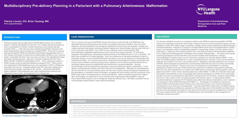
This is a case of a 26 year-old G6P3023 female with asthma, sickle cell trait, and PAVM that was diagnosed between pregnancies during workup for possible pulmonary embolism. Since her initial diagnosis, she had presented to the emergency department several times with dyspnea. Notably, her oxygen saturation was always normal and repeat imaging never demonstrated anything other than her 7 x 6 mm left lower lobe PAVM. Prior CTAP for other indications did not reveal hepatic AVM. Echocardiogram suggested intrapulmonary shunt, but the shunt was not graded. CT pulmonary angiogram described poorly characterized feeding arteries to the PAVM around 2-3 mm in diameter. Our team was consulted at 34 weeks gestation. Given our preference for neuraxial anesthesia for labor or Caesarean section, we recommended MRI brain and spine to rule out the presence of intracerebral and subarachnoid AVMs. Our concerns were risk for intracranial hemorrhage with valsalva during labor and so as to avoid conflation with post dural puncture headache, and to prevent spinal hematoma due to accidental needle trauma to possible AVM while performing a neuraxial anesthetic. We also recommended involving interventional radiology to discuss possible pre-delivery embolization. A pre-delivery multidisciplinary meeting was held to discuss her management. Although her echocardiogram did not comment on her shunt severity, given she was never hypoxic during her dyspneic episodes and her hemoglobin or hematocrit were not elevated, we agreed her shunt fraction and the overall size of the PAVM were small or inconsequential to warrant embolization. When the patient presented to triage in labor at 38 weeks, we made sure to have a double lumen endotracheal tube available in case we encountered PAVM rupture so as to emergently isolate the affected lung. Ultimately, the patient had an uncomplicated vaginal delivery under epidural anesthesia.
Our primary anesthetic concerns in a peripartum patient with PAVM included the possibility of PAVM rupture and subsequent pulmonary hemorrhage, intrapulmonary shunt and subsequent hypoxia, and the possibility of other HHT related organ involvement, namely a lower lumbar subarachnoid AVM precluding neuraxial anesthesia. Pregnancy is thought to increase PAVM volume due to increased plasma, cardiac output, and angiogenic factors, predisposing patients to hemorrhage, stroke, and high cardiac output heart failure 4. Experts in HHT formed a panel in 2011 and 2020 to produce the International Guidelines for the Diagnosis and Management of Hereditary Hemorrhagic Telangiectasia 1. This panel recommends screening all pregnant woman with HHT in the second trimester with transthoracic contrast echocardiography (“bubble study”) and CT pulmonary angiogram for clinically relevant PAVM. In this case, the feeding arteries evaluated on CT were considered small (ie, less than 3 mm), and the patient’s dyspnea was thought to be due to concomitant asthma. Regarding neuraxial anesthesia in the context of PAVM, pharmacological sympathectomy could decrease afterload and worsen shunting and thereby induce dyspnea and hypoxia. However, this was not considered a contraindication to neuraxial anesthesia compared to the risk of hemothorax with mechanical ventilation under general anesthesia. With this case, early identification of a patient with PAVM poses an actionable clinical question, does the patient have HHT; in other words, does the patient have other AVMs? In 1985, Drs. Nicholas Hirsch and Christopher Child describe a case of paraplegia after epidural anesthesia in a patient with a spinal AVM thought to be due to local blood flow disturbance causing spinal cord ischemia 5. However, spinal AVMs that occur in the context of HHT are largely located in the thoracic spine 3. In retrospective studies, neuraxial anesthesia in peripartum HHT patients were performed without complications, even in patients not screened for spinal AVM beforehand with MRI/CT 2,3. Knowledge of the absence of spinal and cerebral AVMs can streamline clinical thinking when encountering much more common postpartum neuropathies or post dural puncture headaches. If lumbar spinal AVM is discovered, the patient should still be offered epidural anesthesia as opposed to CSE and a discussion of the benefits and risks should be held, and if proceeding with epidural, every effort should made to avoid the vertebral level involved. This case underscores the importance of multidisciplinary care coordination in the management of medically complex parturients.
1. Annals of Internal Medicine. (2020). Second International Guidelines for the Diagnosis and Management of Hereditary Hemorrhagic Telangiectasia. https://doi.org/10.7326/M20-1443.
2. Shovlin CL, Sodhi V, McCarthy A, Lasjaunias P, Jackson JE, Sheppard MN. Estimates of maternal risks of pregnancy for women with hereditary haemorrhagic telangiectasia (Osler-Weber-Rendu syndrome): suggested approach for obstetric services. BJOG. 2008 Aug;115(9):1108-15. doi: 10.1111/j.1471-0528.2008.01786.x. Epub 2008 May 30. PMID: 18518871.
3. de Gussem EM, Lausman AY, Beder AJ, Edwards CP, Blanker MH, Terbrugge KG, Mager JJ, Faughnan ME. Outcomes of pregnancy in women with hereditary hemorrhagic telangiectasia. Obstet Gynecol. 2014 Mar;123(3):514-520. doi: 10.1097/AOG.0000000000000120. PMID: 24499751.
4. Shimohira M, Kawai T, Ohta K. An Update on Embolization for Pulmonary Arteriovenous Malformations. Interv Radiol (Higashimatsuyama). 2023 Jun 3;8(2):56-63. doi: 10.22575/interventionalradiology. 2021-0030. PMID: 37485484; PMCID: PMC10359166.