334 posters, 125 sessions, 831 authors, 236 institutions
ePostersLive by SciGen Technologies S.A. All rights reserved.
78th Annual PostGraduate Assembly in Anesthesiology (PGA78) 2024
December 6 - 9, 2024 | New York City

3238
Lumbosacral Plexopathy in the Postpartum State
Part of Topic
Case Title: Lumbosacral Plexopathy in the Postpartum State?
Authors: Mitaire Erhieyovwe MS4., Shivam Patel MS4, Alesha Sands Foster DO3, Larry Lee Jr. MD
Abstract: This case presentation is about a 19 year obstetrics patient who was observed to have a labor complication status post vaginal delivery. Diagnosed while inpatient with lumbosacral plexopathy with mild bilateral obturator nerve neuropraxia and left lumbosacral plexus neuropraxia, she has a rare complication that is stated to happen in only 0.3-2% of the population status post vaginal birth. The assessment consisted of Neurology and Anesthesia ruling out possible fractures, dislocations, extraspinal soft tissue abnormalities, and complications of lumbar epidural catheter placement. With improvement at discharge, the patient was referred to Physical Medicine & Rehabilitation for continued ambulation assistance and physical therapy where her confirmed diagnosis was Femoral Neuropathy in the postpartum state.
Introduction: According to the Anesthesia Patient Safety Foundation, Postpartum Nerve Injuries occur in 0.3-2% of all vaginal deliveries [12]. The majority of nerve injuries are caused by a prolonged second stage of labor, secondary to compression and/or stretching of lower extremity nerves during delivery. The second stage of labor is an important part of labor and what happens during this stage plays a vital role in what will happen postpartum to the mother and baby [5]. The American College of Obstetricians and Gynecologists defines a prolonged second stage as more than 54 minutes for women who are primiparous, 19 minutes for women who are multiparous. 25 minutes should be added for women who have epidural anesthesia [1,4]. During postpartum assessment for nerve damage, anesthetic causes for injury must be evaluated in a timely manner. Although rare, delivery associated nerve injuries are serious and may affect the mother’s care for her newborn. It is important that Obstetricians and Anesthesiologists work together to ensure that patients are treated in a timely manner in order to prevent long term or even permanent nerve damage.
Case Presentation:
A 19 year old G2P1011 patient who presented to labor and delivery at 39 weeks and 3 days with complaints of painful uterine contractions and Premature Rupture of Membranes. She attested to normal fetal movements and denied vaginal bleeding. Her prenatal care was complicated by history of one spontaneous abortion, depression, and history of chlamydia early in pregnancy with a negative test of cure. The patient presented with category 1 fetal monitor tracing and three contractions within 10 minutes on the tocodynamometer. Prior to augmentation of labor using pitocin, the patient requested an epidural for pain management. A lumbar epidural was placed without complication and the patient was determined to be stable by the anesthesia team. The patient proceeded to have a normal spontaneous vaginal delivery.
On postpartum day zero, the epidural infusion was discontinued and the epidural catheter was removed without difficulty. Prior to the patient’s trial of ambulation with nursing staff, she complained of bilateral lower extremity weakness with the left side worse than the right. The patient fell onto her knees when attempting to walk on her own and reported difficulty with ambulation stating, “I cannot move my left knee, it feels numb.” The anesthesia team was called to bedside for further assessment. On exam, knee flexion and extension as well as dorsiflexion and plantar flexion of the ankles were preserved bilaterally with active and passive tension. Limited right hip flexion was noted. Strength was graded 4/5 bilaterally in the lower extremities and sensation was intact.The patient was advised to slowly attempt ambulation with assistance from staff.
The patient was re-examined by anesthesia on postpartum day one and reported improvement with complaints limited to “very slight” numbness over her left anterior knee and left hip weakness. She denied lower back pain or difficulty controlling bowel and bladder functions. A focused physical exam revealed no sensory deficits to cold on the torso, bilateral hips, bilateral anterior and posterior thighs, anterior/posterior/medial/lateral aspects of lower legs or bilateral dorsal and plantar surfaces of the feet. Motor strength was graded 5/5 in bilateral knee extension and flexion, 5/5 bilateral ankle dorsiflexion and plantar flexion, 5/5 bilateral great toe dorsiflexion and plantar extension. However, 4-5/5 in right hip flexion and 2-3/5 in left hip flexion.
On postpartum day two, the neurology team was consulted. During her neurology assessment, the patient endorsed bilateral medial thigh numbness and tingling, not present during her initial examination by anesthesia. The neurology team noted diffuse sensory impairment with pinpoint touch and temperature on the left lower extremity compared to the right. She had no complaints of extrapyramidal symptoms.
The patient was able to ambulate with assistance. Her physical exam was significant for improvement in left hip flexion. Initially, the patient was unable to lift her heel during a straight leg test, but on postpartum day two, she was able to achieve a two inch heel clearance from the bed. When re-examined by the anesthesia team later that day, physical exam findings displayed no signs of epidural hematoma or abscess including fever, chills, severe back pain and bowel or bladder changes.
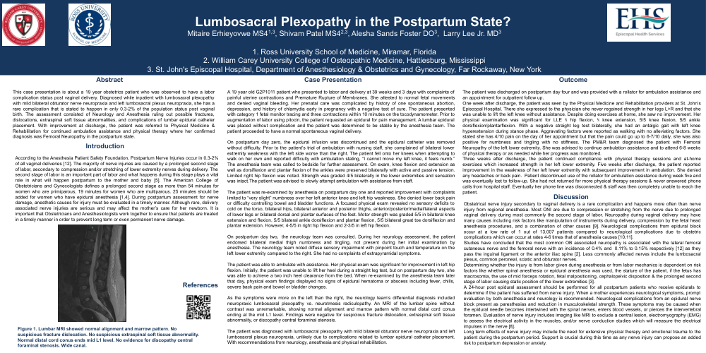
As the symptoms were more on the left than the right, the neurology team’s differential diagnosis included neuropraxic lumbosacral plexopathy vs. neurotmesis radiculopathy. An MRI of the lumbar spine without contrast was unremarkable, showing normal alignment and marrow pattern with normal distal cord conus ending at the mid L1 level. Findings were negative for suspicious fracture dislocation, extraspinal soft tissue abnormality, or discopathy central foraminal stenosis.
The patient was diagnosed with lumbosacral plexopathy with mild bilateral obturator nerve neuropraxia and left lumbosacral plexus neuropraxia, unlikely due to complications related to lumbar epidural catheter placement. With recommendations from neurology, anesthesia and physical rehabilitation.
Outcome:
The patient was discharged on postpartum day four and was provided with a rollator for ambulation assistance and an appointment for outpatient follow up. One week after discharge, the patient was seen by the Physical Medicine and Rehabilitation providers at St. John’s Episocopal Hospital. There she expressed to the physician she never regained strength in her legs L>R and that she was unable to lift the left knee without assistance. Despite doing exercises at home, she saw no improvement. Her physical examination was significant for LLE ⅗ hip flexion, ⅖ knee extension, 5/5 knee flexion, 5/5 ankle dorsiflexion/plantarflexion. With a negative straight leg test bilaterally, she had an antalgic gait with left knee hyperextension during stance phase. Aggravating factors were reported as walking with no alleviating factors. She stated she has 4/10 pain on the day of her appointment but that the pain could go up to 6-7/10 daily, she was also positive for numbness and tingling with no stiffness. The PM&R team diagnosed the patient with Femoral Neuropathy of the left lower extremity. She was advised to continue ambulation assistance and to attend 6-8 weeks of physical therapy or as needed while her progress was monitored.
Three weeks after discharge, the patient continued compliance with physical therapy sessions and at-home exercises which increased strength in her left lower extremity. Five weeks after discharge, the patient reported improvement in the weakness of her left lower extremity with subsequent improvement in ambulation. She denied any headaches or back pain. Patient discontinued use of the rollator for ambulation assistance during week five and was eventually lost to followup. She had not returned for more physical therapy sessions & never answered phone calls from hospital staff. Eventually her phone line was disconnected & staff was then completely unable to reach the patient.
Discussion:
Obstetrical nerve injury secondary to vaginal delivery is a rare complication and happens more often than nerve injury from regional anesthesia. Most ONI are due to compression or stretching from the nerve due to prolonged vaginal delivery during most commonly the second stage of labor. Neuropathy during vaginal delivery may have many causes including risk factors like manipulation of instruments during delivery, compression by the fetal head anesthesia procedures, and a combination of other causes [9]. Neurological complications from epidural block occur at a low rate of 1 out of 13,007 patients compared to neurological complications due to obstetric complications which can occur at rates 4-6 times that of anesthesia causes [10,11].
Studies have concluded that the most common OB associated neuropathy is associated with the lateral femoral cutaneous nerve and the femoral nerve with an incidence of 0.4% and 0.11% to 0.15% respectively [12] as they pass the inguinal ligament or the anterior iliac spine [2]. Less commonly affected nerves include the lumbosacral plexus, common peroneal, sciatic and obturator nerves.
Determining whether the injury is from labor given during anesthesia or from labor mechanics is dependent on risk factors like whether spinal anesthesia or epidural anesthesia was used, the stature of the patient, if the fetus has macrosomia, the use of mid forceps rotation, fetal malpositioning, cephalopelvic disposition & the prolonged second stage of labor causing static position of the lower extremities [3].
A 24-hour post epidural assessment should be performed for all postpartum patients who receive epidurals to determine if the patient has suffered from nerve injury. When a mother experiences neurological symptoms, prompt evaluation by both anesthesia and neurology is recommended. Neurological complications from an epidural nerve block present as paresthesias and reduction in musculoskeletal strength. These symptoms may be caused when the epidural needle becomes intertwined with the spinal nerves, enters blood vessels, or pierces the intervertebral foramen. Evaluation of nerve injury includes imaging like MRI to exclude a central lesion, electromyography (EMG) to assess the electrical activity in the muscles, and/or nerve conduction studies which will measure the electrical impulses in the nerve [8].
Long term effects of nerve injury may include the need for extensive physical therapy and emotional trauma to the patient during the postpartum period. Support is crucial during this time as any nerve injury can propose an added risk to postpartum depression or anxiety.
Summary:
Obstetrical nerve injuries are rare but when they do happen by at least 24 hours postpartum all patients should have evaluations to ensure they do not have any nerve injury to ensure safety for the mother and baby. Methods to prevent labor associated nerve injuries include proper labor positioning, frequent lower extremity repositioning, avoidance of hip hyperflexion during labor and ultimately cesarean section for following deliveries. When labor associated nerve injuries occur, management includes interdisciplinary intervention from anesthesia, neurology, and physical medicine and rehabilitation. Improved function can be expected between 6-8 weeks, with full recovery ranging from 2 months to 24 months.