334 posters, 125 sessions, 831 authors, 236 institutions
ePostersLive by SciGen Technologies S.A. All rights reserved.
78th Annual PostGraduate Assembly in Anesthesiology (PGA78) 2024
December 6 - 9, 2024 | New York City

3236
Anesthetic Management of a Patient with Limb-Girdle Muscular Dystrophy Undergoing an Upper Endoscopy Exam Under General Anesthesia
Poster Presenter
Authors
Affiliations
Part of Topic
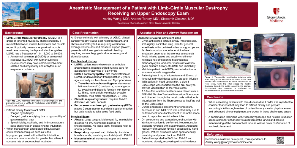
Anesthetic Management of a Patient with Limb-Girdle Muscular Dystrophy Receiving an Upper Endoscopy Exam
Ashley Wang, MD , Andrew Torpey, MD , Slawomir Oleszak, MD; Department of Anesthesiology, Stony Brook University Hospital
Background
• Limb-Girdle Muscular Dystrophy (LGMD) is a group of inherited myopathy characterized by a mismatch between muscle breakdown and muscle repair. It typically presents as proximal muscle weakness involving the hip and shoulder girdles. LGMD has a frequency of 1 in 15,000 to 50,000.
- Autosomal dominant (LGMD1) or autosomal recessive (LGMD2) with further subtypes
- Severe cases may have cardiac involvement (dilated cardiomyopathy and arrhythmia) or respiratory problems.
• Difficult airway features of LGMD
- Tongue hypertrophy
- Delayed gastric emptying due to hypomotility of gastrointestinal tract
- Spinal rigidity, scoliosis, and limb contractures pose challenges in positioning for intubation
• When managing an anticipated difficult airway, combination techniques such as video laryngoscopy paired with a flexible intubation scope are recommended for greater first attempt success rate of endotracheal intubation.
Case Presentation
• A 19-year-old male with a history of LGMD, dilated cardiomyopathy status post heart transplant, and chronic respiratory failure requiring continuous average volume-assured pressure support (AVAPS) presents with lower gastrointestinal bleeding requiring an esophagogastroduodenoscopy and sigmoidoscopy.
• Past Medical History
- LGMD: patient uses wheelchair to ambulate around home, requires skilled nursing care for assistance for activities of daily living
- Dilated cardiomyopathy: rare manifestation of LGMD, underwent heart transplantation 7 years ago, currently on Tacrolimus and Mycophenolate
- Transthoracic echocardiogram: decreased left ventricular (LV) cavity size, normal global LV systolic and diastolic function with normal LV filling, normal right ventricular systolic function, mild mitral regurgitation, EF 60%
- Chronic respiratory failure: requires AVAPS delivered via nasal cannula
- Percutaneous endoscopic gastrostomy (PEG): placed 2 years ago for nutritional supplementation but patient able to take some food orally
• Physical Exam
- Airway: Large tongue, Mallampati IV, Interincisive distance >3 cm, hyomental distance >3 fingerbreadths, No loose teeth, Mobilizes neck to neutral position
- Respiratory: symmetrical, bilaterally diminished breath sounds, breathing comfortably with AVAPS
- Musculoskeletal: contracted upper and lower extremities
• Given anticipated difficult airway (macroglossia, limb rigidity, aspiration risk), plan for general anesthesia with combined video laryngoscope and flexible intubation scope for endotracheal intubation under total intravenous anesthesia
• Avoid inhaled gases and succinylcholine to minimize risk of triggering hyperkalemia, rhabdomyolysis, and other muscular toxicities given patient’s history of LGMD and immobility
• Maintain patient on AVAPS prior to induction to ensure adequate oxygenation
• Patient given 2 mg of midazolam and 50 mcg of fentanyl in divided doses with a propofol infusion
• With patient in head-up position, a size 3 GlideScope was inserted into the oral cavity to provide visualization of the vocal cords
• A 6.0 cuffed oral tracheal tube was placed over a BFP-190 Flexible Tracheal Intubation Fiberscope and directed through the vocal cords with double visualization from the fiberoptic scope itself as well as the GlideScope
• On emergence and extubation, oral suction with Yankauer suction tip performed. Neuromuscular blockade reversed with sugammadex, Adequate recovery of muscular function assessed by hand grasps. Patient extubated while spontaneously breathing and placed back on AVAPS.