334 posters, 125 sessions, 831 authors, 236 institutions
ePostersLive by SciGen Technologies S.A. All rights reserved.
78th Annual PostGraduate Assembly in Anesthesiology (PGA78) 2024
December 6 - 9, 2024 | New York City

3220
A Case of SVC Syndrome Requiring Interdisciplinary Coordination to Optimize Patient Safety
Poster Presenter
Authors
Affiliations
Part of Topic
A Case of SVC Syndrome Requiring Interdisciplinary Coordination to Optimize Patient Safety
Katherine Street BSN, William Chafin BSN, Christopher Schild MD, Judith L. P. Handley MD
Anesthesiology and Perioperative Medicine, Medical College of Georgia, Augusta University, Augusta, Georgia
Introduction:
Superior vena cava (SVC) syndrome occurs due to obstruction of blood flow in the vessel and is frequently caused by an underlying malignancy (65%), but may also be due to benign causes including thrombosis of pacemakers or intravascular catheters. Nonsquamous and squamous cell lung cancers account for 50% and 25% of cases of SVC syndrome respectively. SVC obstruction leads to elevated intravenous pressures in the upper body, resulting in such signs and symptoms as headache, vision changes, reduced level of consciousness, orthopnea, laryngeal edema, epistaxis, and facial edema. Chest computed tomography (CT) with contrast is typically used to identify the obstruction location as well as the underlying cause. Tissue diagnosis is used to plan therapeutic approaches in cases of suspected malignancy. Treatment options include radiation therapy, chemotherapy, and endovascular stenting. Response to radiation may be seen within days to one month (if responsive). Similarly, in chemosensitive tumors, response may be seen within one to two weeks of treatment initiation. Endovascular stenting is classically reserved for severe or refractory symptoms, though earlier stenting is being studied. SVC syndrome can confer significant anesthetic risks, including complete airway obstruction and cardiovascular collapse.
Clinical History:
A 62-year-old male with a history of stage IV squamous cell lung cancer with bone and brain metastases and SVC syndrome presented to the outpatient endoscopy suite for an urgent esophagogastroduodenoscopy (EGD) due to ongoing anemia. The patient had also had a pulmonary embolus on oral anticoagulation.
Case Description:
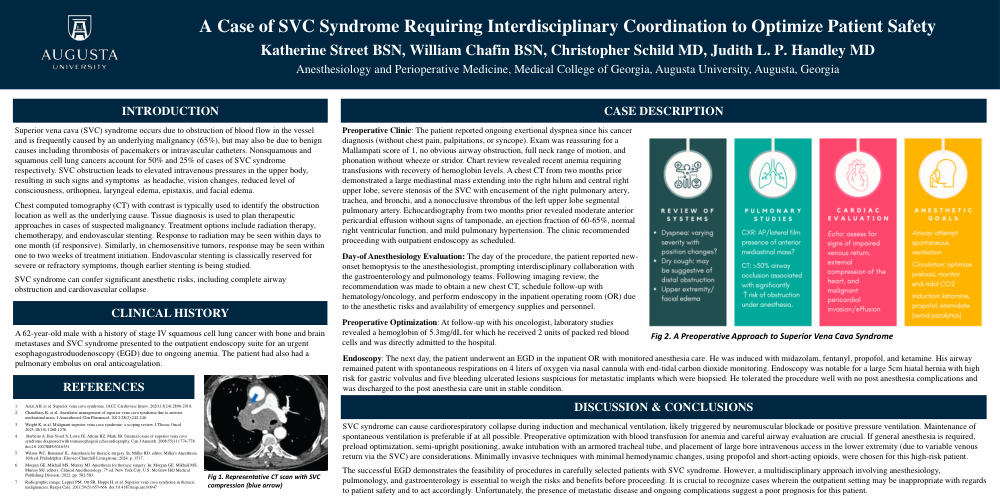
Preoperative Clinic: The patient reported ongoing exertional dyspnea since his cancer diagnosis (without chest pain, palpitations, or syncope). Exam was reassuring for a Mallampati score of 1, no obvious airway obstruction, full neck range of motion, and phonation without wheeze or stridor. Chart review revealed recent anemia requiring transfusions with recovery of hemoglobin levels. A chest CT from two months prior demonstrated a large mediastinal mass extending into the right hilum and central right upper lobe, severe stenosis of the SVC with encasement of the right pulmonary artery, trachea, and bronchi, and a nonocclusive thrombus of the left upper lobe segmental pulmonary artery. Echocardiography from two months prior revealed moderate anterior pericardial effusion without signs of tamponade, an ejection fraction of 60-65%, normal right ventricular function, and mild pulmonary hypertension. The clinic recommended proceeding with outpatient endoscopy as scheduled.
Day-of Anesthesiology Evaluation: The day of the procedure, the patient reported new-onset hemoptysis to the anesthesiologist, prompting interdisciplinary collaboration with the gastroenterology and pulmonology teams. Following imaging review, the recommendation was made to obtain a new chest CT, schedule follow-up with hematology/oncology, and perform endoscopy in the inpatient operating room (OR) due to the anesthetic risks and availability of emergency supplies and personnel.
Preoperative Optimization: At follow-up with his oncologist, laboratory studies revealed a hemoglobin of 5.3mg/dL for which he received 2 units of packed red blood cells and was directly admitted to the hospital.
Endoscopy: The next day, the patient underwent an EGD in the inpatient OR with monitored anesthesia care. He was induced with midazolam, fentanyl, propofol, and ketamine. His airway remained patent with spontaneous respirations on 4 liters of oxygen via nasal cannula with end-tidal carbon dioxide monitoring. Endoscopy was notable for a large 5cm hiatal hernia with high risk for gastric volvulus and five bleeding ulcerated lesions suspicious for metastatic implants which were biopsied. He tolerated the procedure well with no post anesthesia complications and was discharged to the post anesthesia care unit in stable condition.
Discussion & Conclusions:
SVC syndrome can cause cardiorespiratory collapse during induction and mechanical ventilation, likely triggered by neuromuscular blockade or positive pressure ventilation. Maintenance of spontaneous ventilation is preferable if at all possible. Preoperative optimization with blood transfusion for anemia and careful airway evaluation are crucial. If general anesthesia is required, preload optimization, semi-upright positioning, awake intubation with an armored tracheal tube, and placement of large bore intravenous access in the lower extremity (due to variable venous return via the SVC) are considerations. Minimally invasive techniques with minimal hemodynamic changes, using propofol and short-acting opioids, were chosen for this high-risk patient.
The successful EGD demonstrates the feasibility of procedures in carefully selected patients with SVC syndrome. However, a multidisciplinary approach involving anesthesiology, pulmonology, and gastroenterology is essential to weigh the risks and benefits before proceeding. It is crucial to recognize cases wherein the outpatient setting may be inappropriate with regards to patient safety and to act accordingly. Unfortunately, the presence of metastatic disease and ongoing complications suggest a poor prognosis for this patient.
References: