334 posters, 125 sessions, 831 authors, 236 institutions
ePostersLive by SciGen Technologies S.A. All rights reserved.
78th Annual PostGraduate Assembly in Anesthesiology (PGA78) 2024
December 6 - 9, 2024 | New York City

3209
A case of spontaneous spinal cerebrospinal fluid leak with intracranial hypotension, treated by epidural blood patch
Poster Presenter
Authors
Affiliations
Part of Topic
A Case of Spontaneous Intracranial Hypotension, Treated by Epidural Blood Patch
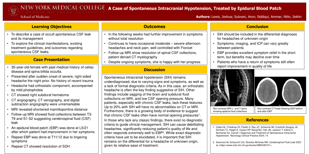
A Case of Spontaneous Intracranial Hypotension, Treated by Epidural Blood Patch • SIH should be included in the differential diagnoses for headaches of unknown origin • Symptoms, imaging, and ICP can vary greatly between patients • EBP provides excellent symptom relief in the short term, but benefits may decline over time • Patients who have a return of symptoms still often report improvement in quality of life Spontaneous intracranial hypotension (SIH) remains underdiagnosed, due to varying signs and symptoms, as well as a lack of formal diagnostic criteria. As in this case, an orthostatic headache is often the key finding suggestive of SIH. Other findings include sagging of the brain and subdural fluid collections on MRI, and low CSF opening pressure. Many patients, especially with chronic CSF leaks, lack these features. Up to 20% with SIH will have no abnormalities on CT or MRI. Furthermore, there is a growing body of evidence to suggest that chronic CSF leaks often have normal opening pressures1. In those who lack any classic findings, there exist no diagnostic criteria to guide clinical management. SIH can cause debilitating headaches, significantly reducing patient’s quality of life and often responds extremely well to EBP2. While exact diagnostic criteria have yet to be elucidated, it is important that SIH remains on the differential for a headache of unknown origin, given its relative ease of treatment. • 35-year-old female with past medical history of celiac disease and spina bifida occulta. • Presented after sudden onset of severe, right sided headache the night prior. No history of recent trauma • Headache had orthostatic component, accompanied by mild photophobia • CT showed right subdural hematoma • CT angiography, CT venography, and digital subtraction angiography were unremarkable • MRI revealed shortened mamillopontinedistance • Follow-up MRI showed fluid collections between T3 T9 and S1-S2 suggesting cerebrospinal fluid (CSF) leak • An epidural blood patch (EBP) was done at L5/21 after which patient had improvement in her symptoms • Repeat EBP was done at T11/12 due to lingering symptoms • Repeat CT showed resolution of SDH • To describe a case of occult spontaneous CSF leak and its management • To explore the clinical manifestations, existing treatment guidelines, and outcomes regarding spontaneous CSF leaks. Learning Objectives 1. Callen AL, Friedman DI, Parikh S, Rau JC, Schievink WI, Cutsforth-Gregory JK, Amrhein TJ, Haight E, Cowan RP, Barad MJ, Hah JM, Jackson T, Deline C, Buchanan AJ, Carroll I: Diagnosis and Treatment of Spontaneous Intracranial Hypotension. Neurology Clinical Practice 2024; 14 2. Severson M, Schaurich CG, Strecker-McGraw MK: Cerebrospinal Fluid Leak 2023 at Outcomes Discussion • In the following weeks had further improvement in symptoms without total resolution • Continues to have occasional moderate – severe afternoon headaches and neck pain, well controlled with Fioricet • Follow-up MRI show resolution of spinal CSF collections, patient denied CT myelography • Despite ongoing symptoms, she is happy with her progress Case Presentation Conclusion References Non-contrast MRI L- and T-spine showing epidural fluid collections Non-contrast CT-head showing SDH before and after EBP Authors: Lewis, Joshua; Sulovari, Aron; Siddiqui, Ammar; Nitin, Sekhri