334 posters, 125 sessions, 831 authors, 236 institutions
ePostersLive by SciGen Technologies S.A. All rights reserved.
78th Annual PostGraduate Assembly in Anesthesiology (PGA78) 2024
December 6 - 9, 2024 | New York City

3162
Aortic Puncture During Transcatheter Mitral Valve in Valve Replacement
Poster Presenter
Authors
Affiliations
Part of Topic
Aortic Puncture During Transcatheter Mitral Valve in Valve Replacement
Introduction:
- Mitral Valve Dysplasia Syndrome (MVDS) is a rare fetal congenital disorder characterized by a collection of multiple cardiac malformations [1,2].
- Typically presents with mitral valve dysplasia, a hypoplastic orifice, and short, thickened chordae tendineae [1,2].
- Can be accompanied by multiple cardiac anomalies, such as a hypoplastic aorta and stenotic aortic valves [1-3].
- Patients also often undergo multiple cardiac surgeries, leading to the formation of scar tissue around the heart.
- Most patients are diagnosed early in life and undergo successful mitral valve replacement
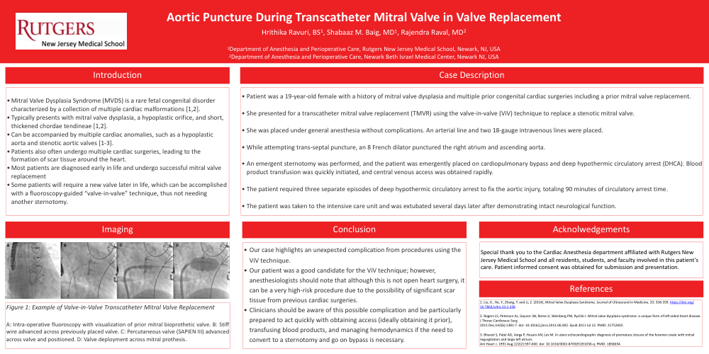
- Some patients will require a new valve later in life, which can be accomplished with a fluoroscopy-guided “valve-in-valve” technique, thus not needing another sternotomy.
Case Description:
- Patient was a 19-year-old female with a history of mitral valve dysplasia and multiple prior congenital cardiac surgeries including a prior mitral valve replacement.
- She presented for a transcatheter mitral valve replacement (TMVR) using the valve-in-valve (ViV) technique to replace a stenotic mitral valve.
- She was placed under general anesthesia without complications. An arterial line and two 18-gauge intravenous lines were placed.
- While attempting trans-septal puncture, an 8 French dilator punctured the right atrium and ascending aorta.
- An emergent sternotomy was performed, and the patient was emergently placed on cardiopulmonary bypass and deep hypothermic circulatory arrest (DHCA). Blood product transfusion was quickly initiated, and central venous access was obtained rapidly.
- The patient required three separate episodes of deep hypothermic circulatory arrest to fix the aortic injury, totaling 90 minutes of circulatory arrest time.
- The patient was taken to the intensive care unit and was extubated several days later after demonstrating intact neurological function.
Conclusion:
- Our case highlights an unexpected complication from procedures using the ViV technique.
- Our patient was a good candidate for the ViV technique; however, anesthesiologists should note that although this is not open heart surgery, it can be a very high-risk procedure due to the possibility of significant scar tissue from previous cardiac surgeries.
- Clinicians should be aware of this possible complication and be particularly prepared to act quickly with obtaining access (ideally obtaining it prior), transfusing blood products, and managing hemodynamics if the need to convert to a sternotomy and go on bypass is necessary.