136 posters, 2 topics, 27 sessions, 339 authors, 88 institutions
ePostersLive by SciGen Technologies S.A. All rights reserved.
2024 ACP Annual Session
NOVEMBER 6-9, 2024 | MARCO ISLAND, FL JW MARRIOT

164
Digital wax up and surgical planning for a patient with Dentinogenesis imperfecta.
Poster Presenter
Authors
Affiliations
DUMMY, University of North Carolina Adams School of Dentistry
Part of Topic
Case Presentation
Digital wax-up and surgical planning for a patient with Dentinogenesis imperfecta.
Amber Olson, DMD, Amara Abreu, DDS, MSD, FACP.
University of North Carolina Adams School of Dentistry, Chapel Hill, NC
INTRODUCTION
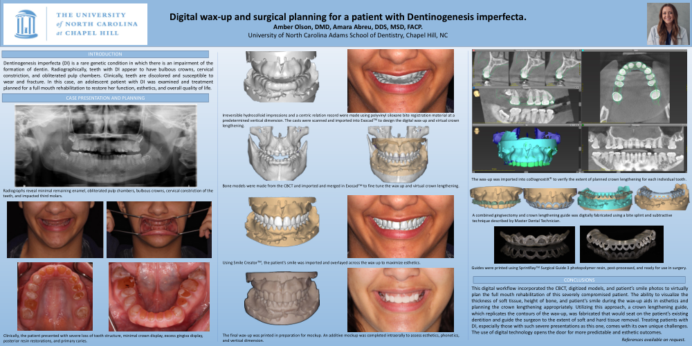
Dentinogenesis imperfecta (DI) is a rare genetic condition in which there is an impairment of the formation of dentin. Radiographically, teeth with DI appear to have bulbous crowns, cervical constriction, and obliterated pulp chambers. Clinically, teeth are discolored and susceptible to wear and fracture. In this case, an adolescent patient with DI was examined and treatment planned for a full mouth rehabilitation to restore her function, esthetics, and overall quality of life.
CASE PRESENTATION AND PLANNING
Radiographs reveal minimal remaining enamel, obliterated pulp chambers, bulbous crowns, cervical constriction of the teeth, and impacted third molars.
Clinically, the patient presented with severe loss of tooth structure, minimal crown display, excess gingiva display, posterior resin restorations, and primary caries.
Irreversible hydrocolloid impressions and a centric relation record were made using polyvinyl siloxane bite registration material at a predetermined vertical dimension. The casts were scanned and imported into ExocadTM to design the digital wax-up and virtual crown lengthening.
Bone models were made from the CBCT and imported and merged in ExocadTM to fine tune the wax up and virtual crown lengthening.
Using Smile CreatorTM, the patient’s smile was imported and overlayed across the wax up to maximize esthetics.
The final wax-up was printed in preparation for mockup. An additive mockup was completed intraorally to assess esthetics, phonetics, and vertical dimension.
The wax-up was imported into coDiagnostiX® to verify the extent of planned crown lengthening for each individual tooth.
A combined gingivectomy and crown lengthening guide was digitally fabricated using a bite splint and Boolean difference technique described by Master Dental Technician.
Guides were printed using SprintRayTM Surgical Guide 3 photopolymer resin, post-processed, and ready for use in surgery.
CONCLUSIONS
This digital workflow incorporated the CBCT, digitized models, and patient’s smile photos to virtually plan the full mouth rehabilitation of this severely compromised patient. The ability to visualize the thickness of soft tissue, height of bone, and patient’s smile during the wax-up aids in esthetics and planning the crown lengthening appropriately. Utilizing this approach, a crown lengthening guide, which replicates the contours of the wax-up, was fabricated that would seat on the patient’s existing dentition and guide the surgeon to the extent of soft and hard tissue removal. Treating patients with DI, especially those with such severe presentations as this one, comes with its own unique challenges. The use of digital technology opens the door for more predictable and esthetic outcomes.