136 posters, 2 topics, 27 sessions, 339 authors, 88 institutions
ePostersLive by SciGen Technologies S.A. All rights reserved.
2024 ACP Annual Session
NOVEMBER 6-9, 2024 | MARCO ISLAND, FL JW MARRIOT

100
Digitally Designed Immediate Denture for a Patient with Failing Implants: A Case Report
Poster Presenter
Authors
Affiliations
Part of Topic
Case Presentation
Combined Workflow For Digitally Designed Immediate Denture in a Patient With Failing Implants: A Case Report
Jawad Tamimi DDS , Rafael Ruiz-Delgado DDS, MSc, PhD, Tanya Somohano-Marquez DMD, FACP
Introduction
This case presentation highlights the utilization of hybrid workflow using digital and conventional methods to create an immediate denture following the implant failure of full arch prosthesis.
Case History
A 64-year-old male patient presented to the postgraduate prosthodontic clinic at Stony Brook University School of Dental Medicine, having been transferred from the care of a previous resident. The patient had a history of multiple implant failures, with no known etiological causes. Surgically guided Straumann SLA Standard Plus Active endosteal implants were placed in sites #20, #22, #27, and #29 in December of 2019. A provisional fixed dental prosthesis (FDP) was placed.
In January of 2022, implants in sites #20 and #29 were removed, and the distal extensions of the FDP were sectioned. Thus, temporarily supporting the sectioned prosthesis. Subsequently, in August of 2024, implants at sites #22 and #27 were also removed, and an immediate mandibular denture was placed on the same day.
Initial Digital Steps
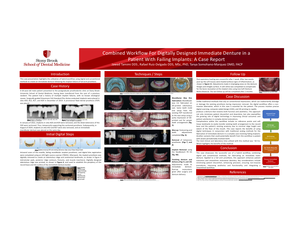
Intraoral scans of the maxilla, failing mandibular implant prosthesis, and digital bite registration were completed using an LED light source scanner (TRIOS). Afterward, the implant prosthesis was digitally removed to create an edentulous ridge and anatomical landmarks, as shown in figure 3 (retromolar pads, posterior ridge contours, frenums, and muscle insertions). Digitally designed edentulous ridge was printed, as shown in Figure 4, and used to establish the periphery of the record base and the outline of the immediate denture border.
Techniques / Steps
Mandibular Wax Rim Fabrication: Mandibular wax rim fabricated on 3D-printed edentulous cast. Same teeth mold and setup from the existing FDP transferred to the wax setup using a putty impression of 3D-printed cast for proper teeth arrangement (fig. 5).
Wax-up: Festooning and teeth adjustments completed (fig. 6).
Removal of temporary prostheses. (Figs 7, and 8)
Implant Removal using the NeoBiotech FR Kit (fig. 8d) .
Finishing denture and delivery (Figs 9, and 10)
Adjustments made to immediate denture. Post-op instructions given after surgery and denture delivery.
Follow Up
Post-operatory healing was uneventful after 1 week. After two weeks post-op the soft tissues were healed without signs of inflammation, as shown in figure 11. Adjustments made in impinging areas on the denture flanges and intaglio surface. A soft reline was completed to compensate for the bone resorption at the implant sites using Coe-Soft Denture Reline Material. The last follow-up will be completed after 2 months.
Case Discussion
Unlike traditional methods that rely on conventional impressions, which can inadvertently dislodge or damage the existing prosthesis during impression removal, the digital workflow offers a non-invasive alternative, which in this case is essential for the patient. The process involves precise digital scanning, computer-aided design (CAD), and 3D printing to rapidly
produce a denture that restores both function and aesthetics. This approach not only minimizes patient discomfort and downtime, but also exemplifies the growing role of digital technology in improving clinical outcomes and patient satisfaction in complex dental restorations.
Complications within this workflow include no reference points and soft tissue landmarks to easily transfer existing teeth arrangement to the record base and the patient’s existing anatomy due to minimal ridge height and extent of the floor of the mouth. This case reports the benefits of using digital techniques in conjunction with traditional analog methods for the fabrication of immediate denture in a complex case involving failing implants. Another scenario that could potentially benefit from this workflow is patient with severe periodontally involved teeth.
The total clinical and laboratory time utilized with this method was ~20 hrs. Which highlights the benefits of this method.
Conclusion
This case showcases the successful use of a hybrid workflow, combining digital and conventional methods, for fabricating an immediate lower denture. Applied to a full arch prosthesis, this approach enhances patient outcomes and streamlines restorative dentistry. Key considerations include minimizing patient discomfort, enhancing precision, ensuring non-invasive procedures, improving aesthetics and functionality, and integrating a streamlined workflow.