136 posters, 2 topics, 27 sessions, 339 authors, 88 institutions
ePostersLive by SciGen Technologies S.A. All rights reserved.
2024 ACP Annual Session
NOVEMBER 6-9, 2024 | MARCO ISLAND, FL JW MARRIOT

10
Ultrasound Assisted Implant Placement for Immediate Temporization
Poster Presenter
Authors
Ebrahim Dastouri, Hsun-Liang Chan, Oliver Kripfgans, Chao-Ho Chien
Affiliations
Part of Topic
Case Presentation
Ultrasound Assisted Implant Placement for Immediate Temporization
Ebrahim Dastouri, D.M.D , Oliver Kripfgans, PhD, Hsun-Liang Chan, DDS, MS, MSD, Chao-Ho Chien, DDS, DScD
Post Graduate Prosthodontics, Department of Restorative Dentistry
Rutgers School of Dental Medicine, Newark, New Jersey, USGA
Introduction
Minimally invasive implant surgery can reduce post-operative morbidity, undesirable tissue volume changes, and mucosal recession. The Palatal Approach Flap (PAF) technique is an ideal flap design for achieving this by maintaining the soft and hard tissues on the buccal side of the alveolar ridge, thereby enhancing aesthetic outcomes and mitigating unpredictable complications. However, this method has limitations, such as inadequate access to the buccal side of the alveolar ridge, which hampers direct visualization. Therefore, imaging assistance during osteotomy is beneficial due to limited access. This study aimed to describe the application of novel ultrasonography (USG) in chairside evaluation for guided minimally invasive implant placement with immediate temporization, enhancing peri-implant hard and soft tissue outcomes.
Material & Method
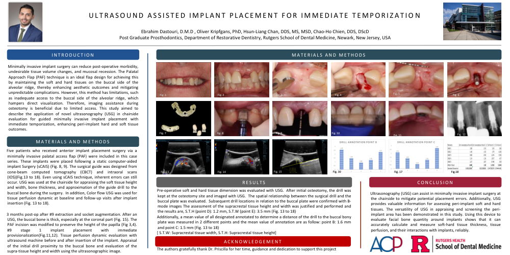
Five patients who received anterior implant placement surgery via a minimally invasive palatal access flap (PAF) were included in this case series. These implants were placed following a static computer-aided implant placement (sCAIP). The surgical guide was designed from cone-beam computed tomography (CBCT) and intraoral scans (IOS). Even using sCAIP technique, inherent errors can still occur. USG was used at the chairside for appraising the soft tissue height and width, bone thickness, and approximation of the guide drill to the buccal bone during the surgery. In addition, Color flow USG was used for tissue perfusion dynamic at baseline and follow-up visits after implant insertion.
3 months post-op after #9 extraction and socket augmentation. After an USG, the buccal bone is thick, especially at the coronal part. The PAF incision was modified to preserve the height of the papilla. #9 stage 1 implant placement with immediate provisionalization. Tissue perfusion dynamic evaluation with ultrasound machine before and after insertion of the implant. Appraisal of the initial drill proximity to the buccal bone and evaluation of the supra-tissue height and width using the ultrasonographic image.
Results
Pre-operative soft and hard tissue dimension was evaluated with USG. After initial osteotomy, the drill was kept at the osteotomy site and imaged with USG. The spatial relationship between the surgical drill and the buccal plate was evaluated. Subsequent drill locations in relation to the buccal plate were confirmed with B-mode images.
Conclusion
Ultrasonography (USG) can assist in minimally invasive implant surgery at the chairside to mitigate potential placement errors. Additionally, USG provides valuable information for assessing peri-implant soft and hard tissues. The versatility of USG in appraising and screening the peri-implant area has been demonstrated in this study. Using this device to evaluate facial bone quantity around implants shows that it can accurately calculate and measure soft-hard tissue thickness, tissue perfusion, and their interactions with implants, reliably.
Acknowledgment
The authors gratefully thank Dr. Priscilla