136 posters, 2 topics, 27 sessions, 339 authors, 88 institutions
ePostersLive by SciGen Technologies S.A. All rights reserved.
2024 ACP Annual Session
NOVEMBER 6-9, 2024 | MARCO ISLAND, FL JW MARRIOT

140
Transforming an Esthetically Challenging Case: A Case Report on Using Innovative Digital Strategies and Solutions
Poster Presenter
Authors
Salma Elsaadany, Mostafa Shehabeldin, Hasan Al Yousuf, Muhammed Saleh, Berna Saglik
Affiliations
Part of Topic
Case Presentation
Introduction:
Implant rehabilitation can be particularly challenging in young adult patients with partial edentulism and advanced periodontitis, especially when it involves the anterior region. Utilizing innovative digital technologies can enhance precision and aid in achieving predictable and superior esthetic results. This clinical case report describes an interdisciplinary, complete digital workflow combining guided implant placement, photogrammetry, 3D-printed prototypes, and digital smile design.
Case Presentation:
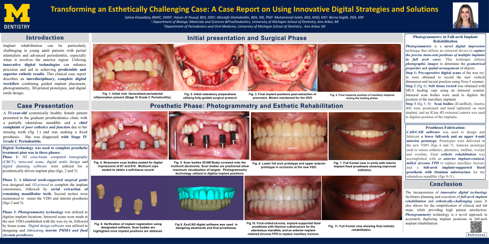
A 33-year-old systemically healthy female patient presented to the graduate prosthodontics clinic with a partially edentulous mandible and a chief complaint of poor esthetics and function due to her missing teeth (fig 1.) and was seeking a fixed prosthesis. She was diagnosed with Stage IV Grade C Periodontitis.
Digital Technology was used to complete prosthetic treatment plan was in three phases:
Phase 1: 3D cone-beam computed tomography (CBCT), intra-oral scans, digital smile design and digital planning software were utilized for a prosthetically-driven implant plan (figs. 2 and 3).
Phase 2: A bilateral tooth-supported surgical guide was designed and 3D-printed to complete the implant osteotomies, followed by serial extraction of remaining mandibular teeth. Second molars were maintained to retain the VDO and interim prosthesis (figs 2 and 3).
Phase 3: Photogrammetry technology was utilized to digitize implant locations. Intraoral scans were made at the new VDO established with the wax try-in, followed by tissue scans. Digital design software was utilized in designing and fabricating interim PMMA and final zirconia prostheses.
Photogrammetry in Full-arch Implant Rehabilitation
Photogrammetry is a novel digital impression technique that utilizes an extraoral device to capture the precise intra-oral positions of multiple implants in full arch cases. This technique utilizes photographic images to determine the geometrical properties and spatial arrangement of objects.
Step 1: Pre-operative digital scans of the wax try-in were obtained to record the new vertical dimension and incisal position using Trios 5 scanner.
Step 2 (fig 4): Soft tissue record was obtained with MUA healing caps using an intraoral scanner. Intraoral scan bodies were used to digitize the position of the maxillary implants
Step 3 (fig 5, 8): Scan bodies (ICamBody; Imetric 4D) were positioned and hand tightened on each implant, and an ICam 4D extraoral camera was used to digitize position of the implants.
Prostheses Fabrication:
CAD/CAM software was used to design and fabricate a lower full-arch and an upper 4-unit anterior prototype. Prototypes were delivered at the new VDO (figs 6 and 7). Anterior prototype used to assess esthetics, phonetics, midline, overjet and overbite. Final esthetic rehabilitation was accomplished with an anterior implant-retained, milled zirconia FPD to replace maxillary incisors and a full-arch implant-supported fixed prosthesis with titanium substructure for the edentulous mandible (figs 9-11).
Conclusion:
The incorporation of innovative digital technology facilitates planning and execution of full-arch implant rehabilitation and esthetically-challenging cases. It also allows for the simplification of clinical and lab steps, while providing high patient satisfaction. Photogrammetry technology is a novel approach in accurately digitizing implant positions in full-arch implant rehabilitation.