136 posters, 2 topics, 27 sessions, 339 authors, 88 institutions
ePostersLive by SciGen Technologies S.A. All rights reserved.
2024 ACP Annual Session
NOVEMBER 6-9, 2024 | MARCO ISLAND, FL JW MARRIOT

14
Virtual Patient-Assisted, Completely Digital Workflow for Fully Guided Maxillary Full-Arch Rehabilitation with Immediate Implant Placement and Loading: A Case Report
Poster Presenter
Authors
Affiliations
Part of Topic
Case Presentation
Virtual Patient-assisted Digital Workflow for Fully Guided Maxillary Full Arch Rehabilitation with Immediate Implant Placement and Loading: A Case Report
Objectives:
Full arch implant rehabilitation requires precise treatment planning and seamless collaboration between surgical and prosthetic teams. The virtual patient workflow offer significant benefit to this process. This case report aims to describe the digital workflow for virtual patient-assisted full arch rehabilitation with implant-supported fixed dental prosthesis.
Introduction:
Recent technological advances have increased the use of digital tools like CBCT, intraoral, and extraoral scanners in dentistry. These tools are integrated into diagnostics and treatment, allowing for the use of CAD/CAM technology to fabricate precise prostheses and implant templates. The virtual patient workflow creates a simulated patient by superimposing 3D images of the patient's face, teeth, and oral structures. This process reduces the number of required appointments while maintaining high-quality digital dental implant surgery planning and prosthetic design.
Case Report:
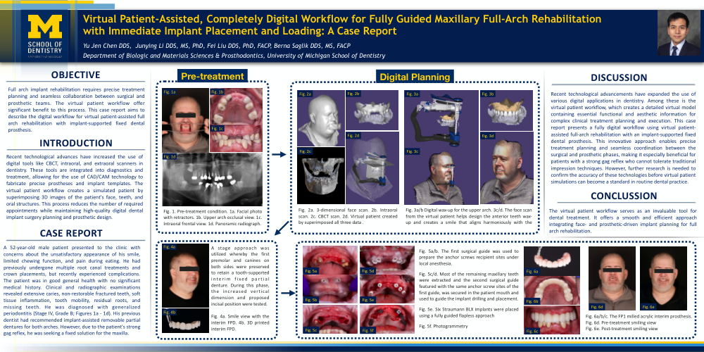
T A 52-year-old male patient presented to the clinic with concerns about the unsatisfactory appearance of his smile, limited chewing function, and pain during eating. He had previously undergone multiple root canal treatments and crown placements, but recently experienced complications. The patient was in good general health with no significant medical history. Clinical and radiographic examinations revealed extensive caries, non-restorable fractured teeth, soft tissue inflammation, tooth mobility, residual roots, and missing teeth. He was diagnosed with generalized periodontitis (Stage IV, Grade B; Figures 3a and 3b). His previous dentist had recommended implant-assisted removable partial dentures for both arches. However, due to the patient's strong gag reflex, he was seeking a fixed solution for the maxilla.
After discussing the risks, limitations, and benefits of different treatment options, the patient chose to proceed with the upper implant-supported fixed complete denture with immediate implant placement and loading. The 3-dimensional facial scans, intraoral scans, and CBCT images of the hard tissue were superimposed using dental CAD software to create a virtual patient. This information is essential for analyzing the smile, achieving harmony with the lips and face, and guiding a 3D digital smile design for complex prosthodontic rehabilitation. A stage approach was utilized whereby the first premolar and canines on both sides were preserved to retain a tooth-supported interim fixed partial denture. Based on the virtual patient, prostheses for immediate loading were designed, and the occlusal scheme was tested in provisional restorations prior to implant.
On the day of the upper arch surgery, the first surgical guide was used to prepare the anchor screws recipient sites under local anesthesia (Figures 8a and 8b). Then, most of the remaining maxillary teeth were extracted and the second surgical guide featured with the same anchor screw sites of the first guide, was secured in the patient mouth and used to guide the implant drilling and placement (Figures 8c and 8d). Six implants (Straumann BLX, Roxolid SLActive; Straumann) were placed in a fully guided flapless approach with an insertion torque of 35 Ncm. All five implants achieved primary stability of more 35 Ncm except for implant #6. Abutments (RW/WB screw-retained abutments; Straumann) were placed under the torque of 35 Ncm for implant #3, 7, 10, 11 and 14, and cover screw placed on implant #6.
A total of three scans were performed on the day of surgery for the fabrication of the full-arch implant-supported fixed prosthesis. First, the interim FDP was digitally scanned using an intraoral scanner (TRIOS, 3Shape A/S, Copenhagen, Denmark). Then, the provisional FDP was removed, and a digital scan of the soft tissue was taken. Finally, scan bodies (iCam4D, iMetric4D Imaging, Courgenay, Switzerland) were placed and scanned extraorally using a photogrammetry scanner after implant placement. The FP1 milled acrylic interim prosthesis was delivered to the patient within 48 hours of the implant surgery, requiring minimal adjustments. The patient was very satisfied with the outcome and returned for checkups at 1 week, 3 weeks, and 1 month, with no biological or prosthetic complications reported.
Discussion:
Recent technological advancements have expanded the use of various digital applications in dentistry. Among these is the virtual patient workflow, which creates a detailed virtual model containing essential functional and aesthetic information for complex clinical treatment planning and execution. This case report presents a fully digital workflow using virtual patient-assisted full-arch rehabilitation with an implant-supported fixed dental prosthesis. This innovative approach enables precise treatment planning and seamless coordination between the surgical and prosthetic phases, making it especially beneficial for patients with a strong gag reflex who cannot tolerate traditional impression techniques. However, further research is needed to confirm the accuracy of these technologies before virtual patient simulations can become a standard in routine dental practice.
Conclusion:
The virtual patient workflow serves as an invaluable tool for dental treatment. It offers a smooth and efficient approach integrating face- and prosthetic-driven implant planning for full arch rehabilitation.