127 posters, 10 topics, 20 sessions, 127 authors
ePostersLive by SciGen Technologies S.A. All rights reserved.
UCSF 2024 Inquiry Symposium
Conference ePosters

P127
Development and Implementation of a 3D-Printed Pulmonary Artery Model for Vascular and Interventional Radiology Simulation Training
Poster Presenter
Authors
Part of Topic
Education Science
Title:
Development and Implementation of a 3D-Printed Pulmonary Artery Model for Vascular and Interventional Radiology Simulation Training
Authors:
Samuel A. Vydro, Scott Drapeau, Adam Alyafaie, Ryan P. Lokken, and Miles B. Conrad
Introduction:
Pulmonary artery (PA) interventions are complex procedures that include pulmonary angiography, thrombectomy, arteriovenous malformation embolization, and balloon angioplasty for chronic thromboembolic pulmonary hypertension. These procedures are an essential skill for interventional radiologists, yet endovascular curriculum is not standardized across accredited fellowships nationwide. Procedural training in interventional radiology (IR) is primarily taught through the Master-Apprentice model. Given the challenging nature of PA interventions and the relative scarcity of procedures per trainee, this methodology invites the potential of patient harm. Simulation training eliminates patient risk in the learning process and has been shown across specialties to improve the speed and quality of skill acquisition as opposed to classical textbook education. However, there are currently no prevalent modalities used for PA simulation.
Hypotheses:
IR trainees will voice subjective improvement in self-confidence regarding conducting PA procedures after training with the model. IR trainees will see a significant reduction in PA access time, fluoroscopy time, and kerma area product between the initial and final training session with the model.
Aims:
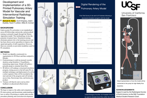
Our aim is to develop a novel, open-source 3D-printed model, incorporating venous, cardiac, and pulmonary arterial components as it relates to endovascular PA interventions to facilitate anatomic and procedural education.
Project Design/Methods:
Using a de-identified, patient CT-scan Digital Imaging and Communications in Medicine file, we isolated the superior vena cava, inferior vena cava, all four heart chambers, and pulmonary arterial tree in 3D-Slicer. The digital model was then sculpted in Blender and finalized in conjunction with the UCSF Maker’s Lab. Primary construction of the final prototype will yield an encased vascular skeleton using StrataSys’s TissueMatrix with efforts to promote clinical fidelity, mimicking organ tissue and allowing for procedural manipulation. Blood flow will be simulated with use of a United Biologics FlowTek 125 pulsatile pump. UCSF IR faculty will assess the prototype via anonymous questionnaires, with adjustments made accordingly.
Efficacy of the model will be evaluated with a series of pre- and post- tests, measuring speed and precision among fellows before and after training with the model. Before the first pre-test, an IR senior faculty member will conduct an introduction to the model, consisting of a detailed explanation of the planned examination technique and demonstration under fluoroscopy. Example tests include timed access to the left main PA with multiple techniques (Omniflush Catheter with stiff Glidewire, Swan-Ganz Catheter, and 6 French Grollman PA Catheter) and time to deploy a coil in a select segment. Data will be recorded as access time, fluoroscopy time, and kerma area product. An extended version of the previous faculty questionnaire will then be distributed to fellows to elicit feedback on educational effectiveness.
Results/Product:
The model will be utilized in a structured simulation program among UCSF IR fellows, simulating intervention. The trainees will submit feedback on its utility in training per distributed surveys. Data regarding trainee feedback and quantitative results of access time, fluoroscopy time, and kerma area product will be gathered and available once the final model prototype is created and utilized in a sample curriculum.
Discussion/Conclusion:
We hope to disseminate understanding of how to utilize 3D printing to enable endovascular procedural planning and curricular development. We believe the final iteration of the model will yield improved competency of PA interventions, and thus patient-safety, once implemented.