145 posters, 12 topics, 25 sessions, 145 authors
ePostersLive by SciGen Technologies S.A. All rights reserved.
UCSF 2023 Inquiry Symposium
10 May | University of San Francisco

P130
Single Nuclear RNA Sequencing of Pituitary Neuroendocrine Tumors Reveals Differences in Tumor Subtype Origin and Development
Poster Presenter
Authors
Part of Topic
Biomedical Science
Introduction:
Pituitary neuroendocrine tumors (PitNETs) are the third most common intracranial tumor in the US. While PitNET subtypes are known to differ by transcription factor staining on pathologic analysis, the developmental heritage and trajectory of these tumors remains unexplored.
Hypothesis:
We hypothesized that PitNET subtypes would originate from different cells of origin in the pituitary gland and behave differently as they develop over time.
Aims:
The aim of this study was to utilize single nuclear RNA sequencing (SnRNA-seq) and trajectory analysis to explore the developmental origins and mitotic trajectories of silent gonadotroph adenomas and prolactinomas, the two most common pituitary neuroendocrine tumors.
Methods:
Tumor samples were collected from patients undergoing endoscopic endonasal resection for pituitary neuroendocrine tumors. Individual cell nuclei were isolated into single nuclear suspension, and each nucleus was tagged with a unique molecular identifier before being sequenced for RNA expression. Tumor nuclei were identified by comparing samples to normal pituitary gland tissue and by exploring copy number variations within each cluster. Single nuclear trajectory analysis was performed where nucleus “pseudotime” was calculated by aggregating differentially expressed genes per cluster relative to pituitary stem cell nuclei. Pseudotime was used to statistically label tumor clusters as “early,” “intermediate,” or “late” in development. Nuclei were further classified by examining cell cycle markers, and statistically different clusters were subsequently labeled as “quiescent” (G0/G1) or “replicating” (G2M/S-Phase). Pseudotime and cell cycle labels were compared to explore mitotic activity over time for PitNET subtypes.
Results/Product:
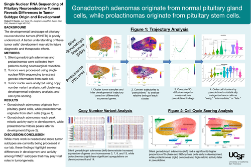
A total of three patient samples (2 silent gonadotroph adenomas, 1 prolactinoma) were processed, resulting in 155,023 sequenced nuclei for analysis. Copy number variant analysis revealed that silent gonadotroph adenomas demonstrate increased expression of genes on chromosomes 3, 7, 8, and 13, while prolactinomas have significant upregulations on chromosomes 8 and 14. Trajectory analysis found that while silent gonadotroph adenomas originate from normal pituitary gland cells, prolactinomas originate from pituitary stem cells instead. Cell cycle cluster labeling revealed that while silent gonadotroph adenomas had a significantly higher proportion of S-phase and G2M-phase cells early in development, prolactinomas demonstrated high mitotic activity late in tumor development.
Discussion/Conclusion:
This study successfully interrogated the lineage and developmental activity of the two most common pituitary neuroendocrine tumor subtypes. We found that while silent gonadotroph adenomas originate from mature pituitary gland cells and reach peak mitotic activity shortly after tumorigenesis, prolactinomas develop from stem cells instead, and are more quiescent until late in tumor development. This finding is surprising given that prolactinomas secrete excess prolactin into the bloodstream and were hypothesized at this study’s inception to develop from lactotroph cells. While this study is currently limited by low sample size and a limited number of PitNET subtypes, we are in the process of sequencing 7 more PitNETs (1 prolactinoma, 3 corticotroph adenomas, 2 somatotroph adenomas, and 1 null cell adenoma), thus bolstering sample diversity and our analytic power. Future directions include analysis of these other PitNETs’ developmental origins and further exploration of differences among tumor subtypes, including cell cycle analysis, gene ontology, and spatial transcriptomics.