2,281 posters, 63 videos, 56 audios, 43 topics, 18 sessions, 6,174 authors, 2,323 institutions
ePostersLive by SciGen Technologies S.A. All rights reserved.
RCOG World Congress 2025
June 23 - 25, 2025 | ExCeL London, UK
EP.1437
Hyperreactio Luteinalis, Pre-Eclampsia and Post-Partum Haemorrhage in a Twin Pregnancy
Poster Presenter
Authors
Affiliations
Part of Topic
Intrapartum care
Hyperreactio Luteinalis, Pre-eclampsia and Postpartum Haemorrhage in a Twin Pregnancy
Objective: This case describes a monochorionic diamniotic (MCDA) twin pregnancy complicated by pre-eclampsia and hyperreactio luteinalis (HL) identified intra-operatively. The delivery was further complicated by a massive post-partum haemorrhage (PPH) of 3.5 liters.
Case Report:
Presentation: A 27-year-old G1P0 woman with MCDA twins presented at 35 weeks gestation to her routine antenatal visit and was found to be hypertensive (BP 140/90mmHg). On examination, she was hyperreflexic bilaterally with clonus and had pitting oedema up to the thighs. She was diagnosed with pre-eclampsia based on examination, blood pressure and a urine protein-creatinine ratio (uPCR) of 38.8 mg/mmol. All other bloods were normal, and no regular anti hypertensives were required antenatally. Delivery was recommended, and she opted for a Caesarean section.
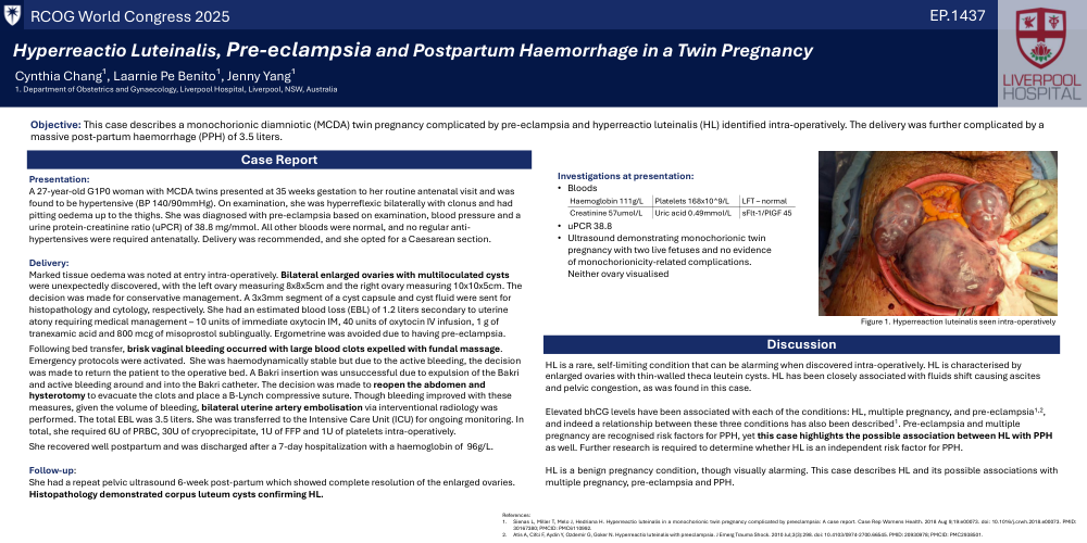
Delivery: Marked tissue oedema was noted at entry intra-operatively. Bilateral enlarged ovaries with multiloculated cysts were unexpectedly discovered, with the left ovary measuring 8x8x5cm and the right ovary measuring 10x10x5cm. The decision was made for conservative management. A 3x3mm segment of a cyst capsule and cyst fluid were sent for histopathology and cytology, respectively. She had an estimated blood loss (EBL) of 1.2 liters secondary to uterine atony requiring medical management – 10 units of immediate oxytocin IM, 40 units of oxytocin IV infusion, 1 g of tranexamic acid and 800 mcg of misoprostol sublingually. Ergometrine was avoided due to having pre-eclampsia. Following bed transfer, brisk vaginal bleeding occurred with large blood clots expelled with fundal massage. Emergency protocols were activated. She was haemodynamically stable but due to the active bleeding, the decision was made to return the patient to the operative bed. A Bakri insertion was unsuccessful due to expulsion of the Bakri and active bleeding around and into the Bakri catheter. The decision was made to reopen the abdomen and hysterotomy to evacuate the clots and place a B-Lynch compressive suture. Though bleeding improved with these measures, given the volume of bleeding, bilateral uterine artery embolisation via interventional radiology was performed. The total EBL was 3.5 liters. She was transferred to the Intensive Care Unit (ICU) for ongoing monitoring. In total, she required 6U of PRBC, 30U of cryoprecipitate, 1U of FFP and 1U of platelets intra-operatively. She recovered well postpartum and was discharged after a 7-day hospitalization with a haemoglobin of 96g/L.
Follow-up: She had a repeat pelvic ultrasound 6-week post-partum which showed complete resolution of the enlarged ovaries. Histopathology demonstrated corpus luteum cysts confirming HL.
Discussion: HL is a rare, self-limiting condition that can be alarming when discovered intra-operatively. HL is characterised by enlarged ovaries with thin-walled theca leutein cysts. HL has been closely associated with fluids shift causing ascites and pelvic congestion, as was found in this case. Elevated bhCG levels have been associated with each of the conditions: HL, multiple pregnancy, and pre-eclampsia, and indeed a relationship between these three conditions has also been described1. Pre-eclampsia and multiple pregnancy are recognised risk factors for PPH, yet this case highlights the possible association between HL with PPH as well. Further research is required to determine whether HL is an independent risk factor for PPH. HL is a benign pregnancy condition, though visually alarming. This case describes HL and its possible associations with multiple pregnancy, pre-eclampsia and PPH