2,281 posters, 63 videos, 56 audios, 43 topics, 18 sessions, 6,174 authors, 2,323 institutions
ePostersLive by SciGen Technologies S.A. All rights reserved.
RCOG World Congress 2025
June 23 - 25, 2025 | ExCeL London, UK
EP.1434
Management of mid-trimester fetal death complicated by complete placenta praevia and pre-eclampsia.
Poster Presenter
Authors
Affiliations
Part of Topic
Intrapartum care
Title: Management of mid-trimester fetal death complicated by complete placenta praevia and pre-eclampsia.
Introduction
Major obstetric haemorrhage (MOH) is recognised as a leading cause of maternal morbidity and mortality. Identifying risk factors, planning, and prompt management are crucial to mitigate adverse effects. Placenta praevia (PP) is typically managed through surgical intervention, however management via vaginal delivery (VD) has been documented. In these cases, managing MOH was a significant concern, and uterine artery embolization (UAE) was shown to be effective.
Intrauterine fetal death (IUFD) after 20 weeks’ gestation in combination with complete placenta previa (CPP) is rare. In this case, pre-eclampsia, which is rarely associated with PP, with an early onset presentation, additionally rare, raised additional risk towards MOH when considering delivery.
Here, the authors report the successful management of IUFD in a pregnancy complicated by CPP and early-onset pre-eclampsia in whom VD was achieved after UAE.
Case Report
A 30-year-old female, SB, P2+0, initially presented at 13+4 weeks’ gestation with recurrent first trimester bleeding, recurrent nausea and vomiting in pregnancy (NVP), and class 1 obesity (BMI 32kgm2). She had a previous Caesarean Section for failure to progress, following a vaginal birth three years prior. Given the risks for fetal growth restriction (FGR), low dose aspirin was initiated. Ms. SB presented again at 19+2 weeks’ gestation with NVP refractory to oral antiemetics and severe right upper quadrant and epigastric pain. There was new onset hypertension and nephrotic range proteinuria (protein: creatinine ratio (PCR) 335.2 mg/mmol). There was persistent transaminitis. An abdominal ultrasound demonstrated a non-obstructing single gallstone in the body of the gallbladder measuring 1.7cm with no pericholecystic fluid and hepatomegaly with no focal abnormalities.
She was diagnosed with early onset pre-eclampsia and biliary colic. Multidisciplinary management included general surgery and gastroenterology. Following an extended inpatient stay, successful management of hypertension occurred alongside thromboprophylaxis. An anomaly scan performed at 23+1 weeks’ gestation demonstrated FGR with an estimated fetal weight of 0.2kg, anhydramnios, absent end diastolic flow of the umbilical artery, and pulmonary hypoplasia. Anterior CPP was noted. All further investigations, including preeclampsia toxaemia screen was within normal limits. Four days later, the umbilical artery flow became reversed. Unfortunately, an IUFD was diagnosed eleven days after, with persistent anterior CPP. She was asymptomatic for labour, haemorrhage or impending eclampsia.
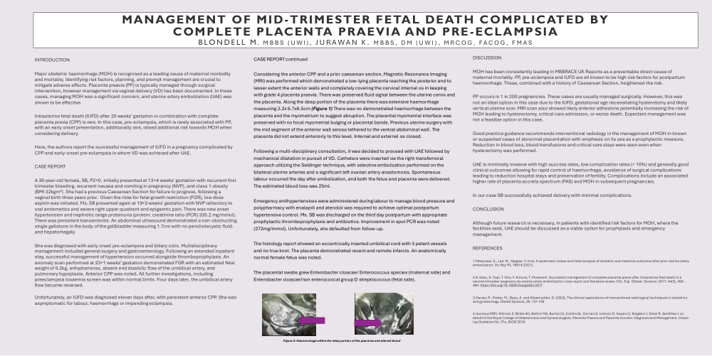
Considering the anterior CPP and a prior caesarean section, Magnetic Resonance Imaging (MRI) was performed which demonstrated a low-lying placenta reaching the posterior and to lesser extent the anterior walls and completely covering the cervical internal os in keeping with grade 4 placenta praevia. There was preserved fluid signal between the uterine cervix and the placenta. Along the deep portion of the placenta there was extensive haemorrhage measuring 2.2x 6.7x6.5cm (Figure 1) There was no demonstrated haemorrhage between the placenta and the myometrium to suggest abruption. The placental myometrial interface was preserved with no focal myometrial bulging or placental bands. Previous uterine surgery with the mid segment of the anterior wall serosa tethered to the ventral abdominal wall. The placenta did not extend anteriorly to this level. Internal and external os closed.
|
|
|
Figure 2: Haemorrhage within the deep portion of the placenta and altered blood.
Following a multi-disciplinary consultation, it was decided to proceed with UAE followed by mechanical dilatation in pursuit of VD. Catheters were inserted via the right transfemoral approach utilizing the Seldinger technique, with selective embolization performed on the bilateral uterine arteries and a significant left ovarian artery anastomosis. Spontaneous labour occurred the day after embolization, and both the fetus and placenta were delivered. The estimated blood loss was 25ml.
Emergency antihypertensives were administered during labour to manage blood pressure and polypharmacy with enalapril and atenolol was required to achieve optimal postpartum hypertensive control. Ms. SB was discharged on the third day postpartum with appropriate prophylactic thromboprophylaxis and antibiotics. Improvement in spot PCR was noted (272mg/mmol). Unfortunately, she defaulted from follow-up.
The histology report showed an eccentrically inserted umbilical cord with 3 patent vessels and no true knot. The placenta demonstrated recent and remote infarcts. An anatomically normal female fetus was noted.
The placental swabs grew Enterobacter cloacae/ Enterococcus species (maternal side) and Enterobacter cloacae/non enterococcal group D streptococcus (fetal side).
Discussion
MOH has been consistently leading in MBBRACE UK Reports as a preventable direct cause of maternal mortality. PP, pre-eclampsia and IUFD are all known to be high risk factors for postpartum haemorrhage. These, combined with a history of Caesarean Section, heightened the risk.
PP occurs in 1 in 200 pregnancies. These cases are usually managed surgically. However, this was not an ideal option in this case due to the IUFD, gestational age necessitating hysterotomy and likely vertical uterine scar. MRI scan also showed likely anterior adhesions potentially increasing the risk of MOH leading to hysterectomy, critical care admission, or worse death. Expectant management was not a feasible option in this case.
Good practice guidance recommends interventional radiology in the management of MOH in known or suspected cases of abnormal placentation with emphasis on its use as a prophylactic measure. Reduction in blood loss, blood transfusions and critical care stays were seen even when hysterectomy was performed.
UAE is minimally invasive with high success rates, low complication rates (< 10%) and generally good clinical outcomes allowing for rapid control of haemorrhage, avoidance of surgical complications leading to reduction hospital stays and preservation of fertility. Complications include an associated higher rate of placenta accreta spectrum (PAS) and MOH in subsequent pregnancies.
In our case SB successfully achieved delivery with minimal complications.
Conclusion
Although future research is necessary, in patients with identified risk factors for MOH, where the facilities exist, UAE should be discussed as a viable option for prophylaxis and emergency management.
References:
1.Matsuzaki, S., Lee, M., Nagase, Y. et al. A systematic review and meta-analysis of obstetric and maternal outcomes after prior uterine artery embolization. Sci Rep 11, 16914 (2021).
2.S. Kaku, S. Tsuji, T. Ono, F. Kimura, T. Murakami. Successful management of complete placenta previa after intrauterine fetal death in a second-trimester pregnancy by uterine artery embolization: case report and literature review. Clin. Exp. Obstet. Gynecol. 2017, 44(3), 458–460. https://doi.org/10.12891/ceog3353.2017
3.Davies, R., Parker, M., Basu, A. and Alleemudder, D. (2023), The clinical applications of interventional radiological techniques in obstetrics and gynaecology. Obstet Gynecol, 25: 131-139
4.Jauniaux ERM, Alfirevic Z, Bhide AG, Belfort MA, Burton GJ, Collins SL, Dornan S, Jurkovic D, Kayem G, Kingdom J, Silver R, Sentilhes L on behalf of the Royal College of Obstetricians and Gynaecologists. Placenta Praevia and Placenta Accreta: Diagnosis and Management. Green-top Guideline No. 27a. BJOG2018