501 posters, 2 videos, 1 audios, 8 topics, 37 sessions, 395 authors, 307 institutions
ePostersLive by SciGen Technologies S.A. All rights reserved.
23rd Annual Pain Medicine Meeting 2024
November 21-23, 2024 | Caesars Palace, Las Vegas, Nevada

6037
ANATOMIC EVALUATION OF SACROILIAC JOINT DIMENSIONS: AN IMAGING SERIES STUDY TO DEFINE SAFETY PARAMETERS
Poster Presenter
Authors
Affiliations
Part of Topic
Scientific Abstracts > Chronic Pain
ANATOMIC EVALUATION OF SACROILIAC JOINT DIMENSIONS:
AN IMAGING SERIES STUDY TO DEFINE SAFETY PARAMETERS
Usman Latif MD, MBA1, Christopher Lam MD1, Sarah Keim PhD2, Nicholas Wharton MS3, Dawood Sayed MD1, Douglas Beall MD4, Timothy Deer MD5
Sacroiliac joint dysfunction is implicated in causing 15-30% of all non-radicular low back pain [1]. Several recent advances in interventional techniques have allowed for minimally invasive posterior fusion of the sacroiliac joint (SIJ). Traditional open and minimally invasive surgery involve a lateral fusion approach whereby an implant is placed from the lateral ilium into the sacrum and is associated with risks of vascular disruption of the superior gluteal artery and injury to the sacral nerves [2, 3]. The anatomic dimensions of the SIJ and the landmarks utilized for lateral implant placement, however, have not been formally studied. By understanding the statistical variations of the anatomy, it may be possible to decrease the incidence of neurovascular complications with lateral SIJ fusions. Here we present an imaging study of salient SIJ parameters to evaluate anatomic dimensions important for surgical planning.
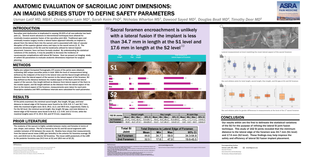
Eighty-one subject Computed Tomography (CT) scans of the pelvis were obtained, evaluating 162 unique sacroiliac joints in total. With the level of measurement being defined as the midpoint of the level in the lateral view and the Sacral length defined as distance from the lateral aspect of the sacrum to the lateral aspect of the foramen, SIJ Gap defined as the distance between the medial aspect of the ilium and the lateral aspect of the sacrum, Iliac length defined as distance from lateral aspect of the ilium to the medial aspect, and SIJ length defined as the distance from the lateral aspect of the ilium to the lateral aspect of the foramen, measurements were taken for each joint. Descriptive statistics and 95% confidence intervals were calculated for each parameter
Of the joints examined, the minimal sacral length, iliac length, SIJ gap, and total distance to lateral edge of S1 foramen were found to be 13.6, 6.9, 1.7 and 34.7 mm, while the maximal lengths were 32.5, 29.2, 11.3, and 59.9 mm, respectively (Table 1). For the S2 level, the minimal sacral length, iliac length, SIJ gap, and total distance to lateral edge of S2 foramen were found to be 8.2, 1.2, 0.00, and 17.6 mm, while the maximal lengths were 27.5, 24.3, 9.8, and 47.9 mm, respectively.
The anatomy of the sacrum is highly variable between males and females in terms of size, shape, and contour. The SIJ is formed by the S1 and S2 sacral segments with variable inclusion of S3 between the sexes [4]. Studies have shown that measurements from the lateral sacral mass (LSM) (par lateralis) to the anterior S1 foramina average 29 mm, and 22.8 mm to the anterior S2 foramina. The mean width posteriorly of the LSM to the posterior foramina was 24.3 mm at S1 and 18.6 mm at S2 [5].
Our results within are the first to delineate the statistical variations of the SIJ for the purpose of refining the lateral SI joint fusion technique. This study of 162 SI joints revealed that the minimum distance to the lateral edge of the foramen was 34.7 mm (S1 level) and 17.6 mm (S2 level). These findings may help improve the safety and efficiency of lateral SIJ fusion implant placement.