501 posters, 2 videos, 1 audios, 8 topics, 37 sessions, 395 authors, 307 institutions
ePostersLive by SciGen Technologies S.A. All rights reserved.
23rd Annual Pain Medicine Meeting 2024
November 21-23, 2024 | Caesars Palace, Las Vegas, Nevada

5665
THE COMPLEX REGIONAL PAIN SYNDROME (CRPS) CONUNDRUM - CLINICAL JUDGMENT VS. PARADOXICAL SCANS: A CASE REPORT
Poster Presenter
Part of Topic
Scientific Abstracts > Acute Pain
The Complex Regional Pain Syndrome (CRPS) Conundrum - Clinical Judgment vs. Paradoxical Scans: A Case Report
Karthik Kamath, Elliott Berry, Leslie Wong, Jenzel Espares, Peter Atallah, Ajendra Sohal Nassau University Medical Center (NUMC)
Introduction
33-year-old male diagnosed with Complex Regional Pain Syndrome (CRPS). The patient, a restrained driver, presented after a motor vehicle collision. He denied loss of consciousness but complained of back, bilateral leg, and right hand/wrist pain. A pan scan revealed a small subdural hemorrhage. After stabilization, he was deemed suitable for acute inpatient rehabilitation. Throughout his admission, he experienced severe right foot/ankle pain with allodynia and hyperalgesia, avoiding weight-bearing. Pain management included gabapentin, duloxetine, and oxycodone. Mild to moderate RLE swelling developed, with negative X-rays for fracture and vascular dopplers for DVT. Meeting Budapest criteria, he was diagnosed with complex regional pain syndrome (CRPS) and discharged home with outpatient PT and pain management follow-up for potential intervention.
Materials and Methods
As the case report is devoid of patient identifiable information, it is exempt from IRB review requirements as per Nassau Univeristy Medical Center (NUMC) policy.
Results/Case Report
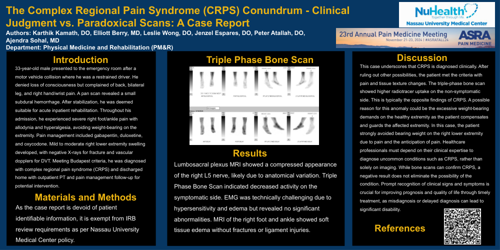
Lumbosacral plexus MRI showed a compressed appearance of the right L5 nerve, likely due to anatomical variation. Triple Phase Bone Scan indicated decreased activity on the symptomatic side. EMG was technically challenging due to hypersensitivity and edema but revealed no significant abnormalities. MRI of the right foot and ankle showed soft tissue edema without fractures or ligament injuries.
Discussion
This case underscores that CRPS is diagnosed clinically. After ruling out other possibilities, the patient met the criteria with pain and tissue texture changes. The triple-phase bone scan showed higher radiotracer uptake on the non-symptomatic side. This is typically the opposite findings of CRPS. A possible reason for this anomaly could be the excessive weight-bearing demands on the healthy extremity as the patient compensates and guards the affected extremity. In this case, the patient strongly avoided bearing weight on the right lower extremity due to pain and the anticipation of pain. Healthcare professionals must depend on their clinical expertise to diagnose uncommon conditions such as CRPS, rather than solely on imaging. While bone scans can confirm CRPS, a negative result does not
eliminate the possibility of the condition. Prompt recognition of clinical signs and symptoms is crucial for improving prognosis and quality of life through timely treatment, as misdiagnosis or delayed diagnosis can lead to significant disability.
References
Bruehl, S, Harden, NR. Galer, BS, et al. Complex regional pain syndrome: are there distinct subtypes and sequential stages of the syndrome?. Pain 2002 95(1):p 119-124. https://doi.org/10.1016/s0304- 3959(01)00387-6
Johnson, S, Cowell, F, Gillespie, S, et al. Complex regional pain syndrome what is the outcome?‐a systematic review of the course and impact of CRPS at 12 months from symptom onset and beyond. European Journal of Pain 26.6 2022: 1203-1220. https://doi.org/10.1002/ejp.1953
Kessler, A, Min Y, Randy C. Complex regional pain syndrome: An updated comprehensive review. NeuroRehabilitation 47.3 2020: 253-264. https://doi.org/10.3233/nre-208001
Mettler Jr, FA, Guiberteau, MJ. Essentials of Nuclear Medicine Imaging. 6th edition. Philadelphia, PA: Elsevier Health Sciences; 2012.
Stanton-Hicks M, Baron R, Boas R, et al. Complex Regional Pain Syndromes: guidelines for therapy. Clin J Pain 1998;14:155-166 https://doi.org/10.1097/00002508-199806000-00012