501 posters, 2 videos, 1 audios, 8 topics, 37 sessions, 395 authors, 307 institutions
ePostersLive by SciGen Technologies S.A. All rights reserved.
23rd Annual Pain Medicine Meeting 2024
November 21-23, 2024 | Caesars Palace, Las Vegas, Nevada

6189
ACCURACY OF ANTEROPOSTERIOR AND CONTRALATERAL OBLIQUE FLUOROSCOPIC VIEWS FOR LUMBAR INTERLAMINAR EPIDURAL STEROID INJECTION
Poster Presenter
Authors
Affiliations
Part of Topic
Scientific Abstracts > Chronic Pain
ACCURACY OF ANTEROPOSTERIOR AND CONTRALATERAL OBLIQUE FLUOROSCOPIC VIEWS FOR LUMBAR INTERLAMINAR EPIDURAL STEROID INJECTION
Afrin Sagir, MD, Jatinder Gill, MD, Nasir Hussain, MD
Introduction
Epidural steroid injection (ESI) is a commonly performed interventional procedure for treating radicular pain. Proceduralists may encounter false loss of resistance while advancing the needle for interlaminar epidural access. To assess the needle depth and trajectory in relation to the bony landmarks in the spine, contralateral oblique and/or lateral fluoroscopic views are often used while performing ESI. Studies have demonstrated 100% accuracy in needle placement within the epidural space when utilizing these additional views. There is ongoing debate about whether the anteroposterior (AP) view alone is sufficient in most cases, with some arguing that further views should only be employed when necessary to avoid unnecessary regulations and additional radiation exposure to the patient.
However, the clinician's ability to distinguish epidural contrast spread from non-epidural spread in the AP view is subjective and untested. This study aims to determine the accuracy of both the AP and contralateral oblique (CLO) views in distinguishing between contrast spread patterns inside and outside the epidural space. The authors hypothesize that the AP view may not reliably differentiate contrast spread inside the epidural space from superficial, non-epidural spread.
Materials and Methods
This single-center study was performed in the outpatient pain clinic of a major academic hospital from March 2022 to July 2023. The study received Institutional Review Board approval before initiation. Twenty-five adult patients aged 18 years and older, undergoing lumbar interlaminar epidural steroid injection, were included. Exclusion criteria encompassed pregnant patients and those with known allergies to iodinated contrast agents. One patient was excluded post-enrollment due to technical difficulties in accessing the epidural space. Written informed consent was obtained from all study subjects before enrollment.
For the procedure, patients were positioned prone, and a 20-gauge Tuohy needle was aseptically advanced using the loss of resistance technique with the AP view. Before entering the epidural space (verified by radiologic imaging displaying the Tuohy needle proximal to the ventral interlaminar line in the CLO view), 0.5-1.0 ml of iodinated contrast dye was injected. Images of the contrast spread in both AP and CLO views were saved. Following entry into the epidural space, another 0.5-1.0 ml of contrast dye was injected, and corresponding images in both AP and CLO views were saved. The procedure adhered to standard protocols dictated by the clinical situation. In cases of false loss of resistance encountered by the proceduralist before the ventral interlaminar line, 0.5-1ml of contrast was injected, and images in AP and CLO views were saved.
A control group of 24 images, each representing true loss of resistance in the epidural space in AP and CLO views with 0.5-1.0 ml of injected contrast dye, was identified through chart review of recent lumbar epidural steroid injections. The images for both the study and control groups were de-identified and presented in a random order to 10 senior interventional proceduralists who had a minimum of 5 years of clinical experience in pain medicine, who were blinded. These clinicians were tasked with determining whether the images depicted true or false loss of resistance.
Sample Size Justification: The survey questionnaire given to the interventionalists featured images showcasing contrast spread, presenting an equal number of patterns depicting both epidural and non-epidural diffusion. In this study, the inclusion of 24 true and 24 false comparisons offers clinically relevant evidence to assess whether additional views are necessary for accurate deposition of medication in the epidural space.
Statistical Analysis: We used Fleiss’ Kappa as the statistical measure to assess the level of agreement among the proceduralists, beyond what would be expected by chance. It ranges from -1 to 1, where 1 indicates perfect agreement, 0 indicates agreement expected by chance, and negative values suggest less agreement than expected by chance. We report Fleiss’ kappa and the overall accuracy for each proceduralist.
Results
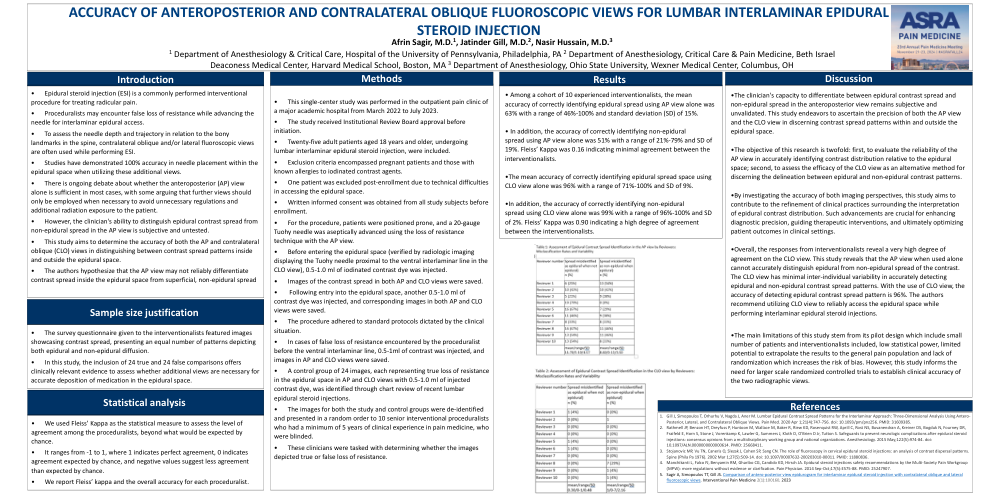
Among a cohort of 10 experienced interventionalists, the mean accuracy of correctly identifying epidural spread using AP view alone was 63% with a range of 46%-100% and standard deviation (SD) of 15%. In addition, the accuracy of correctly identifying non-epidural spread using AP view alone was 51% with a range of 21%-79% and SD of 19%. Fleiss’ Kappa was 0.16 indicating minimal agreement between the interventionalists.
The mean accuracy of correctly identifying epidural spread space using CLO view alone was 96% with a range of 71%-100% and SD of 9%. In addition, the accuracy of correctly identifying non-epidural spread using CLO view alone was 99% with a range of 96%-100% and SD of 2%. Fleiss’ Kappa was 0.90 indicating a high degree of agreement between the interventionalists.
Discussion
The clinician's capacity to differentiate between epidural contrast spread and non-epidural spread in the anteroposterior view remains subjective and unvalidated. This study endeavors to ascertain the precision of both the AP view and the CLO view in discerning contrast spread patterns within and outside the epidural space.
The objective of this research is twofold: first, to evaluate the reliability of the AP view in accurately identifying contrast distribution relative to the epidural space; second, to assess the efficacy of the CLO view as an alternative method for discerning the delineation between epidural and non-epidural contrast patterns.
By investigating the accuracy of both imaging perspectives, this study aims to contribute to the refinement of clinical practices surrounding the interpretation of epidural contrast distribution. Such advancements are crucial for enhancing diagnostic precision, guiding therapeutic interventions, and ultimately optimizing patient outcomes in clinical settings.
Overall, the responses from interventionalists reveal a very high degree of agreement on the CLO view. This study reveals that the AP view when used alone cannot accurately distinguish epidural from non-epidural spread of the contrast. The CLO view has minimal inter-individual variability in accurately detecting epidural and non-epidural contrast spread patterns. With the use of CLO view, the accuracy of detecting epidural contrast spread pattern is 96%. The authors recommend utilizing CLO view to reliably access the epidural space while performing interlaminar epidural steroid injections.
The main limitations of this study stem from its pilot design which include small number of patients and interventionalists included, low statistical power, limited potential to extrapolate the results to the general pain population and lack of randomization which increases the risk of bias. However, this study informs the need for larger scale randomized controlled trials to establish clinical accuracy of the two radiographic views.
Conclusion:
The mean accuracy for correctly identifying epidural spread as epidural and non-epidural spread as non-epidural among 10 experienced interventionalists in the AP view was 63% and 51%. Using the CLO view alone the accuracy increases to 96% and 99% respectively. This study reveals that utilizing standalone AP view without CLO view was inadequate to distinguish epidural from non-epidural spread during lumbar interlaminar epidural access.