501 posters, 2 videos, 1 audios, 8 topics, 37 sessions, 395 authors, 307 institutions
ePostersLive by SciGen Technologies S.A. All rights reserved.
23rd Annual Pain Medicine Meeting 2024
November 21-23, 2024 | Caesars Palace, Las Vegas, Nevada

6006
NEW UPPER EXTREMITY WEAKNESS: ATYPICAL POLYMYALGIA RHEUMATICA VERSUS CERVICAL MYELOPATHY
Poster Presenter
Authors
Affiliations
Part of Topic
Scientific Abstracts > Chronic Pain
Introduction
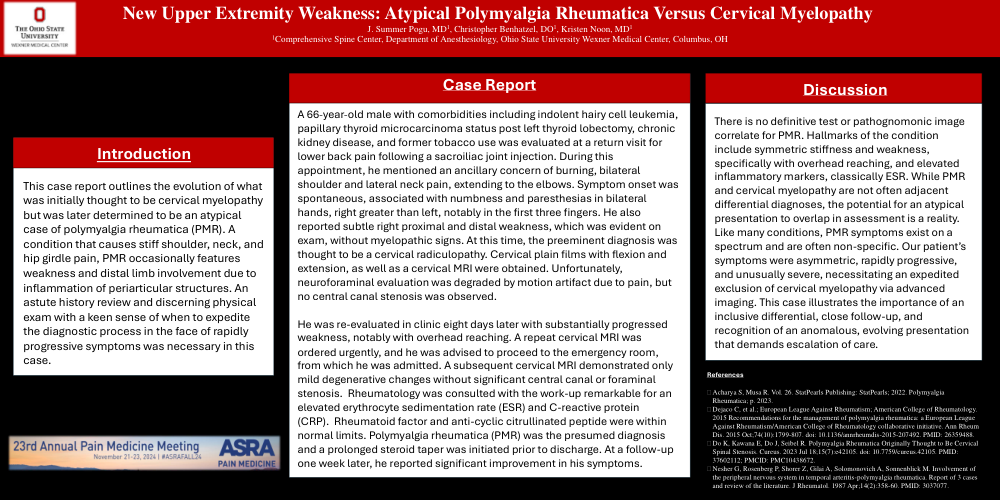
This case report outlines the evolution of what was initially thought to be cervical myelopathy but was later determined to be an atypical case of polymyalgia rheumatica (PMR). A condition that causes stiff shoulder, neck, and hip girdle pain, PMR occasionally features weakness and distal limb involvement due to inflammation of periarticular structures. An astute history review and discerning physical exam with a keen sense of when to expedite the diagnostic process in the face of rapidly progressive symptoms was necessary in this case.
Case Report
A 66-year-old male with comorbidities including indolent hairy cell leukemia, papillary thyroid microcarcinoma status post left thyroid lobectomy, chronic kidney disease, and former tobacco use was evaluated at a return visit for lower back pain following a sacroiliac joint injection. During this appointment, he mentioned an ancillary concern of burning, bilateral shoulder and lateral neck pain, extending to the elbows. Symptom onset was spontaneous, associated with numbness and paresthesias in bilateral hands, right greater than left, notably in the first three fingers. He also reported subtle right proximal and distal weakness, which was evident on exam, without myelopathic signs. At this time, the preeminent diagnosis was thought to be a cervical radiculopathy. Cervical plain films with flexion and extension, as well as a cervical MRI were obtained. Unfortunately, neuroforaminal evaluation was degraded by motion artifact due to pain, but no central canal stenosis was observed.
He was re-evaluated in clinic eight days later with substantially progressed weakness, notably with overhead reaching. A repeat cervical MRI was ordered urgently, and he was advised to proceed to the emergency room, from which he was admitted. A subsequent cervical MRI demonstrated only mild degenerative changes without significant central canal or foraminal stenosis. Rheumatology was consulted with the work-up remarkable for an elevated erythrocyte sedimentation rate (ESR) and C-reactive protein (CRP). Rheumatoid factor and anti-cyclic citrullinated peptide were within normal limits. Polymyalgia rheumatica (PMR) was the presumed diagnosis and a prolonged steroid taper was initiated prior to discharge. At a follow-up one week later, he reported significant improvement in his symptoms.
Discussion
There is no definitive test or pathognomonic image correlate for PMR. Hallmarks of the condition include symmetric stiffness and weakness, specifically with overhead reaching, and elevated inflammatory markers, classically ESR. While PMR and cervical myelopathy are not often adjacent differential diagnoses, the potential for an atypical presentation to overlap in assessment is a reality. Like many conditions, PMR symptoms exist on a spectrum and are often non-specific. Our patient’s symptoms were asymmetric, rapidly progressive, and unusually severe, necessitating an expedited exclusion of cervical myelopathy via advanced imaging. This case illustrates the importance of an inclusive differential, close follow-up, and recognition of an anomalous, evolving presentation that demands escalation of care.