501 posters, 2 videos, 1 audios, 8 topics, 37 sessions, 395 authors, 307 institutions
ePostersLive by SciGen Technologies S.A. All rights reserved.
23rd Annual Pain Medicine Meeting 2024
November 21-23, 2024 | Caesars Palace, Las Vegas, Nevada

5978
STEPPING INTO TROUBLE: FOOT DROP FOLLOWING AN ACETABULAR FRACTURE
Poster Presenter
Authors
Affiliations
Part of Topic
Scientific Abstracts > Acute Pain
Stepping Into Trouble: Foot Drop Following an Acetabular Fracture
53-year-old male sustained a mechanical fall while ambulating across a set of train tracks. He subsequently developed severe pain in his left hip which reportedly radiated distally. He was then transported to the emergency department where imaging then revealed a comminuted and displaced left acetabular fracture extending into the iliac wing and a left superior pubic ramus fracture with minimally displaced left inferior pubic ramus fracture. Imaging was unrevealing of axial injury. He was evaluated by orthopedics and recommended for surgical fixation. He subsequently underwent open reduction and internal fixation of the left acetabulum using the ilioinguinal approach. Post-operatively he continued to experience left lower extremity (LLE) pain initiating in his left hip region with radiation down the posterolateral aspect of his LLE and terminating over the dorsum of his left foot. Pain was described as burning in nature. Additionally, he was noted to have diminished motor strength of the extensor hallucis longus and tibialis anterior.
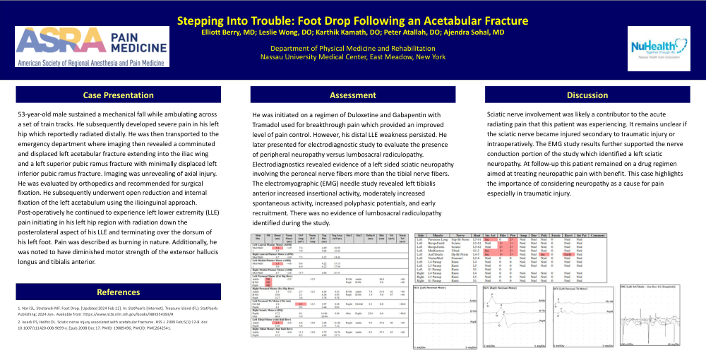
He was initiated on a regimen of Duloxetine and Gabapentin with Tramadol used for breakthrough pain which provided an improved level of pain control. However, his distal LLE weakness persisted. He later presented for electrodiagnostic study to evaluate the presence of peripheral neuropathy versus lumbosacral radiculopathy. Electrodiagnostics revealed evidence of a left sided sciatic neuropathy involving the peroneal nerve fibers more than the tibial nerve fibers. The electromyographic (EMG) needle study revealed left tibialis anterior increased insertional activity, moderately increased spontaneous activity, increased polyphasic potentials, and early recruitment. There was no evidence of lumbosacral radiculopathy identified during the study.
Sciatic nerve involvement was likely a contributor to the acute radiating pain that this patient was experiencing. It remains unclear if the sciatic nerve became injured secondary to traumatic injury or intraoperatively. The EMG study results further supported the nerve conduction portion of the study which identified a left sciatic neuropathy. At follow-up this patient remained on a drug regimen aimed at treating neuropathic pain with benefit. This case highlights the importance of considering neuropathy as a cause for pain especially in traumatic injury.