501 posters, 2 videos, 1 audios, 8 topics, 37 sessions, 395 authors, 307 institutions
ePostersLive by SciGen Technologies S.A. All rights reserved.
23rd Annual Pain Medicine Meeting 2024
November 21-23, 2024 | Caesars Palace, Las Vegas, Nevada

5855
RADIOFREQUENCY ABLATION: PIONEERING THE TREATMENT FRONTIER – A HISTORICAL PERSPECTIVE
Poster Presenter
Part of Topic
Scientific Abstracts > Chronic Pain
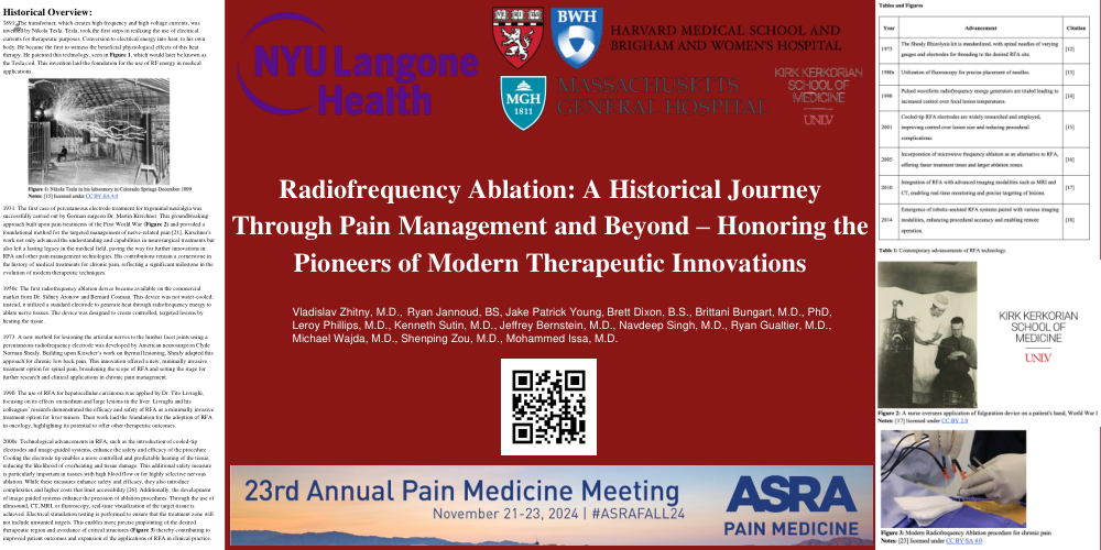
Radiofrequency Ablation: A Historical Journey Through Pain Management and Beyond – Honoring the Pioneers of Modern Therapeutic Innovations
Historical Overview:
1891: The transformer, which creates high frequency and high voltage currents, was invented by Nikola Tesla. Tesla, took the first steps in realizing the use of electrical currents for therapeutic purposes. Conversion to electrical energy into heat, to his own body. He became the first to witness the beneficial physiological effects of this heat therapy. He patented this technology, seen in Figure 1, which would later be known as the Tesla coil. This invention laid the foundation for the use of RF energy in medical applications.
1931: The first case of percutaneous electrode treatment for trigeminal neuralgia was successfully carried out by German surgeon Dr. Martin Kirschner. This groundbreaking approach built upon pain treatments of the First World War (Figure 2) and provided a foundational method for the targeted management of nerve-related pain [21]. Kirschner's work not only advanced the understanding and capabilities in neurosurgical treatments but also left a lasting legacyin the medical field, paving the way for further innovations in RFA and other pain management technologies. His contributions remain a cornerstone in the history of medical treatments for chronic pain, reflecting a significant milestone in the evolution of modern therapeutic techniques.
1950s: The first radiofrequency ablation device became available on the commercial market from Dr. Sidney Aronow and Bernard Cosman. This device was not water-cooled; instead, it utilized a standard electrode to generate heat through radiofrequency energy to ablate nerve tissues. The device was designed to create controlled, targeted lesions by heating the tissue.
1973: A new method for lesioning the articular nerves to the lumbar facet joints using a percutaneous radiofrequency electrode was developed by American neurosurgeon Clyde Norman Shealy. Building upon Kirscher’s work on thermal lesioning, Shealy adapted this approach for chronic low back pain. This innovation offered a new, minimally invasive treatment option for spinal pain, broadening the scope of RFA and setting the stage for further research and clinical applications in chronic pain management.
1990: The use of RFA for hepatocellular carcinoma was applied by Dr. Tito Livraghi, focusing on its effects on medium and large lesions in the liver. Livraghi and his colleagues’ research demonstrated the efficacy and safety of RFA as a minimally invasive treatment option for liver tumors. Their work laid the foundation for the adoption of RFA in oncology, highlighting its potential to offer other therapeutic outcomes.
2000s: Technological advancements in RFA, such as the introduction of cooled-tip electrodes and image-guided systems, enhance the safety and efficacy of the procedure. Cooling the electrode tip enables a more controlled and predictable heating of the tissue, reducing the likelihood of overheating and tissue damage. This additional safety measure is particularly important in tissues with high blood flow or for highly selective nervous ablation. While these measures enhance safety and efficacy, they also introduce complexities and higher costs that limit accessibility [26]. Additionally, the development of image guided systems enhance the precision of ablation procedures. Through the use of ultrasound, CT, MRI, or fluoroscopy, real-time visualization of the target tissue is achieved. Electrical stimulation testing is performed to ensure that the treatment zone will not include unwanted targets. This enables more precise pinpointing of the desired therapeutic region and avoidance of critical structures (Figure 3) thereby contributing to improved patient outcomes and expansion of the applications of RFA in clinical practice.