425 posters, 1 videos, 9 audios, 8 topics, 53 sessions, 340 authors, 286 institutions
ePostersLive by SciGen Technologies S.A. All rights reserved.
22nd Annual Pain Medicine Meeting 2023
November 10 - 11, 2023 | New Orleans Marriott | New Orleans, Louisiana, USA

4786
IMMEDIATE HORNER’S SYNDROME IN A PEDIATRIC PATIENT AFTER PARAVERTEBRAL BLOCK FOR MANAGEMENT OF CHRONIC CHEST PAIN
Poster Presenter
Authors
Affiliations
Part of Topic
Medically Challenging Cases
Pediatric Chest Pain Management with Paravertebral Blocks Resulting in Horner’s Syndrome
Co-Authors:
Naldine Isaac, MD
Kaitlyn Egan, MD
Ravi Dipak Shah, MD
Background
Pectus excavatum is a congenital deformity of the cartilage connecting the ribs to the sternum and results in a depression of the sternum. Symptoms can vary widely and can include shortness of breath, exercise intolerance, fatigue, and chest pain; whereas some cases are asymptomatic. In severe cases there can be compression of the myocardium. Severity often worsens during adolescent growth spurt. Pain management for pectus excavatum is often multimodal including physical therapy, medications, breathing exercises, and lifestyle changes. Nerve blocks can also be an important component of pain management. One option is a paravertebral block of the associated spinal nerves.
Horner’s Syndrome is a neurological condition that results in disruption of the sympathetic nerve pathway. It results in a triad of symptoms: ipsilateral miosis, ptosis, and anhidrosis. Local anesthetics injected near the stellate ganglion can cause a transient Horner’s syndrome. This network of nerve fibers is located on the anterolateral aspect of the neck at the level of the C7 and T1 vertebrae underneath the sternocleidomastoid and scalene muscles.
Case Presentation
A 16 year old right-hand dominant male baseball player with a history of pectus excavatum presented for left chest pain management. Reported pain occurs at rest as well as with exertion, and has been ongoing for the past 2 years. Onset of pain was associated with growth of six inches in height in one year and no other inciting event. Reported constant pain since onset with acute exacerbations approximately every 2 weeks. Exacerbations were accompanied by confusion, lightheadedness/dizziness, and sometimes shortness of breath. Patient’s father noted the patient had failed an exam during one of these episodes; since then, parents elect to keep him home when pain is most severe. He has not discontinued playing baseball as the exacerbations of pain are random and without evidence of initiation by physical activity. The severity of pain episodes has previously necessitated patient evaluation in the Pediatric Emergency department with reported 10/10 on pain scale; he presented with tenderness to palpation of the left pectoralis muscle and intercostal muscles anteriorly below the 2nd rib as well as spasms of left pectoralis and intercostal muscles. No allodynia was noted. Patient had unrestricted range of motion of joints but increase in pain intensity was noted with active or passive movement. Work up included an unremarkable EKG and echocardiogram. CBC, CMP, troponin all were within normal limits. Patient was referred to Pain Management.
The patient previously trialed medication management with gabapentin 100 mg daily progressing to BID, celecoxib 200 mg BID x14 days then PRN, and ibuprofen PRN; none of these conservative treatments provided sufficient relief of pain exacerbations. He is involved in a chronic pain program which includes physical therapy and psychological support. However, he continues to have functionally limiting chest wall pain and thus was scheduled for left paravertebral nerve blocks.
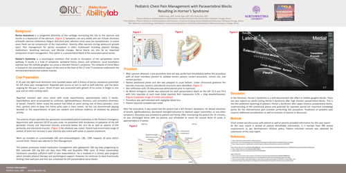
After consent obtained, a pre-procedure time out was performed immediately before the procedure with all team members present to validate correct patient, correct procedure, correct site, and correct position. Patient positioned prone and skin was prepped in usual fashion. Under ultrasound guidance the vascular, muscular, osseous, and pleural structures were identified. Skin infiltration with 1% lidocaine was administered prior to injections. An 80mm echogenic needle was advanced for each paravertebral block on the left T2-3 and T3-4 with 5mL injectate at each level (total injectate 8mL ropivacaine 0.2% + 2mg dexamethasone). Procedure was well tolerated with negligible blood loss. Patient reported complete pain relief. After the procedure, it was noted that the patient had a left Horner's Syndrome. He denied shortness of breath, lightheadedness, decreased strength/sensation to bilateral upper extremities, or any other symptoms. Education was provided to patient and family. After monitoring the patient for 15 minutes, he was discharged home with his parents and scheduled to return for second block of series in approximately 2-3 weeks.
Methods
Chart review and discussion with patient as well as parents provided information for this case report.
As the case report is devoid of patient identifiable information, it is exempt from IRB review requirements as per Northwestern McGaw policy. Patient informed consent was obtained for submission of this case report.
Discussion
In the literature, Horner’s Syndrome is a well-documented side effect in stellate ganglion blocks. There are case reports on adults noting Horner’s Syndrome after high thoracic paravertebral blocks. This is the first published reporting of pediatric Horner’s Syndrome after upper thoracic paravertebral blocks. Understanding of the anatomical planes and potential for injectate spread are important knowledge points for the interventional pain provider performing this procedure. Treatment of pediatric pain requires additional consideration as well as inclusion of parents in discussion.