425 posters, 1 videos, 9 audios, 8 topics, 53 sessions, 340 authors, 286 institutions
ePostersLive by SciGen Technologies S.A. All rights reserved.
22nd Annual Pain Medicine Meeting 2023
November 10 - 11, 2023 | New Orleans Marriott | New Orleans, Louisiana, USA

4500
ULTRASOUND-GUIDED ILIOINGUINAL NERVE RADIOFREQUENCY ABLATION FOR CHRONIC INGUINAL NEURALGIA AFTER FLUOROSCOPY FAILED
Poster Presenter
Authors
Affiliations
Part of Topic
Medically Challenging Cases
RIGHT ilioinguinal/ilio Nerve Radiofrequency Ablation for Ilioinguinal neuralgia Rt.
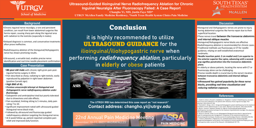
Ultrasound-Guided Ilioinguinal Nerve Radiofrequency Ablation for Chronic Inguinal Neuralgia After Fluoroscopy Failed: A Case Report
Introduction
Chronic inguinal nerve neuralgia is an infrequent, persistent condition that may develop following surgery involving lower abdominal incisions, such as hernia repair 1. This condition commonly manifests as sharp or stabbing pain along the inguinal area, with pain radiating to the testicles in males, significantly impeding the patient's quality of life. Diagnosis of this condition is often delayed, and conservative management options are limited and sometimes ineffective. Radiofrequency ablation of the ilioinguinal/iliohypogastric nerve has been reported to yield favorable outcomes. Ultrasound, a versatile modality, allows for the identification of the ilioinguinal nerve and real-time confirmation of needle placement. This case report highlights a patient who underwent a successful repeated radiofrequency ablation of the ilioinguinal/iliohypogastric nerve under ultrasound guidance after a prior failed attempt under fluoroscopy.
Materials and Methods
The UTRGV Institutional Review Board (IRB) has determined this case report to be exempt from IRB review requirements as it does not constitute research, in accordance with UTRGV policy.
The informed consent for the publication of this case report was obtained from the patient.
Case Report
The patient is an 88-year-old male who presented with chronic right groin pain, characterized as sharp and radiating to the right testicle. These symptoms arose after an inguinal hernia surgery in 2014. The pain was easily reproduced by palpation in the right lower quadrant of the abdomen, and the Carnett sign was positive. The patient had a BMI of 41. A previous pain physician attempted radiofrequency ablation of the ilioinguinal/iliohypogastric nerve under fluoroscopy guidance several years ago, but no improvement was observed. Subsequently, the patient tried gabapentin and amitriptyline for pain control, but these medications were poorly tolerated due to drowsiness and anticholinergic side effects. The pain worsened to the point where the patient could not sit for more than 5 minutes, and he experienced daily pain ranging from 7 to 10 on a numeric rating scale. The patient demonstrated significant improvement with ultrasound-guided ilioinguinal nerve block trials and subsequently underwent ultrasound and fluoroscopy-guided radiofrequency ablation targeting the ilioinguinal nerve. At the 6-week follow-up after the procedure, the patient reported complete pain relief and improved function in daily activities.
Discussion
The ilioinguinal and iliohypogastric nerves are susceptible to injury during abdominal surgeries, such as hernia repair, due to their superficial locations. These nerves run between the transverse abdominis muscle and the internal oblique muscle at the level of the anterior superior iliac spine and then traverse the inguinal canal to become subcutaneous. Ilioinguinal/iliohypogastric nerve blocks have been recognized as effective treatment options for chronic inguinal neuralgia in patients who have not responded to conservative management with GABAergic agents and antiepileptics. However, due to the limited duration of their effectiveness, radiofrequency ablation has been recommended for those experiencing chronic symptoms 2. Traditionally, fluoroscopy or CT has been utilized for needle guidance during the procedure, with successful outcomes reported due to the well-established anatomical landmarks of the ilioinguinal nerve 3. The needle is inserted into the skin at a point 2 cm medial and 2 cm superior to the anterior superior iliac spine and then advanced until a second pop is felt, indicating penetration into the transverse abdominis plane 1. Nevertheless, in elderly or obese patients, locating the nerve solely with CT or fluoroscopy can be challenging. Furthermore, due to the nerve's course between the transverse abdominis and internal oblique muscles, precise needle depth is crucial for the success of nerve block and radiofrequency ablation. Recently, ultrasound has gained popularity for ilioinguinal/iliohypogastric nerve blocks due to its real-time visualization of the target nerve and needle placement 45. Additionally, ultrasound offers the advantage of reduced radiation exposure. In conclusion, the authors recommend considering ultrasound guidance for the treatment of chronic inguinal neuralgia with radiofrequency ablation, particularly in elderly or obese patients for whom the identification of anatomical landmarks for the ilioinguinal/iliohypogastric nerve may be challenging.
Reference
1. Kastler A, Aubry S, Piccand V, Hadjidekov G, Tiberghien F, Kastler B. Radiofrequency neurolysis versus local nerve infiltration in 42 patients with refractory chronic inguinal neuralgia. Pain physician. 2012;15(3):237-244. https://www.ncbi.nlm.nih.gov/pubmed/22622908. doi: 10.36076/ppj.2012/15/237.
2. Chernishof V, Wong J, Kim E. Ilioinguinal/iliohypogastric pulsed radiofrequency for the treatment of pediatric chronic abdominal pain: A case report. A&A Case Reports. 2021;15(2):e01393. https://www.ncbi.nlm.nih.gov/pubmed/33560645. doi: 10.1213/XAA.0000000000001393.
3. Miccio J, Vincent F, Navlani R, Bonder JH, Singh JR. Fluoroscopically-guided pulsed radiofrequency neurotomy technique for the treatment of genitofemoral neuralgia. Pain physician. 2016;19(7):E1099-E1101. https://www.ncbi.nlm.nih.gov/pubmed/27676683. doi: 10.36076/ppj/2016.19.E1099.
4. Demirci A, Efe EM, Türker G, et al. Iliohypogastric/ilioinguinal nerve block in inguinal hernia repair for postoperative pain management: Comparison of the anatomical landmark and ultrasound guided techniques. Brazilian journal of anesthesiology (Elsevier). 2014;64(5):350-356. https://dx.doi.org/10.1016/j.bjane.2014.01.001. doi: 10.1016/j.bjane.2014.01.001.
5. Trainor D, Moeschler S, Pingree M, et al. Landmark-based versus ultrasound-guided ilioinguinal/iliohypogastric nerve blocks in the treatment of chronic postherniorrhaphy groin pain: A retrospective study. Journal of Pain Research. 2015;8(default):767-770. https://www.ncbi.nlm.nih.gov/pubmed/26604817. doi: 10.2147/JPR.S86777.