425 posters, 1 videos, 9 audios, 8 topics, 53 sessions, 340 authors, 286 institutions
ePostersLive by SciGen Technologies S.A. All rights reserved.
22nd Annual Pain Medicine Meeting 2023
November 10 - 11, 2023 | New Orleans Marriott | New Orleans, Louisiana, USA

4701
NOVEL USE OF PULSED RADIOFREQUENCY ABLATION FOR SYMPTOMATIC SURAL NEUROMA IN A TRANSTIBIAL AMPUTEE
Poster Presenter
Authors
Affiliations
Part of Topic
Medically Challenging Cases
Novel Use of Pulsed Radiofrequency Ablation for Symptomatic Sural Neuroma in a Transtibial Amputee
Introduction
Individuals with limb loss are at risk for unique pain conditions not seen in the general population including RLP and phantom limb pain1. Residual limb pain is exceedingly common after amputation and is the most common reason for having to discontinue prosthesis use. Among the most common causes are neuromas, osteophytes and heterotopic ossification, soft tissue friction, and adventitious bursitis2. Of those causes, neuromas have been the most well studied. Within a year of amputation, neuromas will invariably form. The severed nerve will eventually grow back into a neuroma3. While most neuromas do not cause significant pain or symptoms, some do, especially if they are located more superficially, or are prone to pressures or shearing forces in the prosthetic socket. They should be suspected with when direct compression of a focal region of the residual limb causes neuropathic-type pain, especially if in the location one would expect of a lower limb nerve. In some individuals, it will reproduce phantom sensations in the distribution of that nerve. Some guidelines recommend diagnostic ultrasound combined with sonopalpationof the visualized neuroma4.
Conservative treatment of residual limb neuromas, such as medication and limiting prosthesis use, does not address the root cause. However, the literature on percutaneous interventions is lacking. Several small case series and case reports report successful treatment residual limb neuromas using various interventions, including steroid, botulinum toxin, alcohol, or radiofrequency ablation5–8. However, no randomized controlled trials exist. Furthermore, the described interventions exclusively focused on neuromas formed from larger nerves, and not of small, purely sensory nerves. Despite the knowledge that neuromas of small, purely sensory nerve are frequently pain generators in the residual limb (including the sural and saphenous nerves), no literature reports any percutaneous interventions for these. For the sural nerve, there are three case reports that report interventions on the sural nerve in non-amputees, which used cryoablation, pulsed radiofrequency ablation (pRFA), or hydrodissection9–11. In this case report, we describe the treatment of a transtibial amputee’s sural neuroma using pRFA.
Materials and Methods
Written informed consent was obtained from the patient for submission of this case report. As the case report is devoid of patient identifiable information, it is exempt from Institutional Review Board review requirements as per Brooke Army Medical Center policy.
Case History
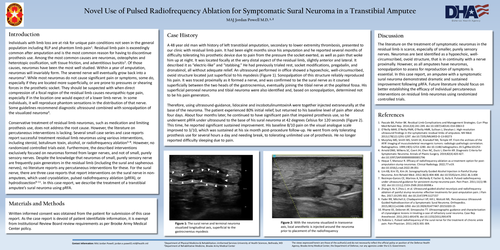
A 48 year oldman with history of left transtibial amputation, secondary to lower extremity thrombosis, presented to our clinic with residual limb pain. It had been eight months since his amputation and he reported several months of difficulty tolerating his prosthetic device due to pain from the pressure the socket exerted, as well as pain that woke him up at night. It was located focally at the very distal aspect of the residual limb, slightly anterior and lateral. It described it as “electric-like” and “stabbing.” He had previously trialed rest, socket modifications, pregabalin, and dronabinol, all without adequate relief. An ultrasound performed in office showed a hypoechoic, well-circumscribed, ovoid structure located just superficial to his myodesis(Figure 1). Sonopalpationof this structure reliably reproduced his pain. It was traced proximally as it formed a nerve, andwas confirmed to be the sural nerve as it coursed superficially between the two heads of the gastrocnemius, eventually joining the tibial nerve at the popliteal fossa. His superficial peroneal neuroma and tibial neuroma were also identified and,based on sonopalpation, determined not to be his pain generators.
Therefore, using ultrasound-guidance, lidocaine and incobotulinumtoxinAwere together injected extraneurallyat the base of the neuroma. The patient experienced 80% initial relief, butreturned to his baseline level of pain after about four days. About four months later, he continued to have significant pain that impaired prosthesis use, so he underwent pRFAunder ultrasound to the base of his sural neuroma at 42 degrees Celsius for 120 seconds (Figure 2). This time, he reported significant sustained improvement. His baseline 8/10 pain on the visual analogue scale improved to 3/10, which was sustained at his six monthpost-procedure follow-up. He went from only tolerating prosthesis use for several hours a day and needing break, to tolerating unlimited use of prosthesis. He no longer reported difficulty sleeping due to pain.
Discussion
The literature on the treatment of symptomatic neuromas in the residual limb is scarce, especially of smaller, purely sensory nerves. Neuromas are best identified as a hypoechoic, well-circumscribed, ovoid structure, that is in continuity with a nerve proximally. However, as all amputees have neuromas, sonopalpationto assess for reproduction of symptoms is essential. In this case report, an amputee with a symptomatic sural neuroma demonstrated dramatic and sustained improvement following pRFA. Future literature should focus on better establishing the efficacy of individual percutaneous interventions on residual limb neuromas using randomized controlled trials.
References
1.Pascale BA, Potter BK. Residual Limb Complications and Management Strategies. CurrPhys Med RehabilRep. 2014;2(4):241-249. doi:10.1007/s40141-014-0063-0
2.O’Reilly MAR, O’Reilly PMR, O’Reilly HMR, Sullivan J, Sheahan J. High-resolution ultrasound findings in the symptomatic residual limbs of amputees. Mil Med. 2013;178(12):1291-1297. doi:10.7205/MILMED-D-13-00273
3.Murphey MD, Smith WS, Smith SE, KransdorfMJ, Temple HT. From the archives of the AFIP. Imaging of musculoskeletal neurogenic tumors: radiologic-pathologic correlation. Radiographics. 1999;19(5):1253-1280. doi:10.1148/radiographics.19.5.g99se101253
4.Arnold DMJ, Wilkens SC, CoertJH, Chen NC, DucicI, EberlinKR. Diagnostic Criteria for Symptomatic Neuroma. Annals of Plastic Surgery. 2019;82(4):420-427. doi:10.1097/SAP.0000000000001796
5.Hoque T, Mansour R. Efficacy of radiofrequency ablation as a treatment option for post-amputation stump neuromas. Clinical Radiology. 2022;77:e19. doi:10.1016/j.crad.2022.09.051
6.Lim KB, Kim YS, Kim JA. SonographicallyGuided Alcohol Injection in Painful Stump Neuroma. Ann RehabilMed. 2012;36(3):404-408. doi:10.5535/arm.2012.36.3.404
7.Restrepo-Garces CE, MarinovA, McHardy P, FaclierG, Avila A. Pulsed radiofrequency under ultrasound guidance for persistent stump-neuroma pain. Pain Pract. 2011;11(1):98-102. doi:10.1111/j.1533-2500.2010.00398.x
8.Zhang X, Xu Y, Zhou J, et al. Ultrasound-guided alcohol neurolysis and radiofrequency ablation of painful stump neuroma: effective treatments for post-amputation pain. J Pain Res. 2017;10:295-302. doi:10.2147/JPR.S127157
9.Fader RR, Mitchell JJ, ChadayammuriVP, Hill J, Wolcott ML. Percutaneous Ultrasound-Guided Hydrodissectionof a Symptomatic Sural Neuroma. Orthopedics. 2015;38(11):e1046-1050. doi:10.3928/01477447-20151020-15
10.RhameEE, DebonetAF, SimopoulosTT. Ultrasonographic guidance and characterization of cryoanalgesiclesions in treating a case of refractory sural neuroma. Case Rep Anesthesiol. 2011;2011:691478. doi:10.1155/2011/691478
11.Todorov L. Pulsed radiofrequency of the sural nerve for the treatment of chronic ankle pain. Pain Physician. 2011;14(3):301-304.