425 posters, 1 videos, 9 audios, 8 topics, 53 sessions, 340 authors, 286 institutions
ePostersLive by SciGen Technologies S.A. All rights reserved.
22nd Annual Pain Medicine Meeting 2023
November 10 - 11, 2023 | New Orleans Marriott | New Orleans, Louisiana, USA

4517
TARGETED PAIN MANAGEMENT AIDED BY ELECTRODIAGNOSIS AFTER TRIMALLEOLAR ANKLE FRACTURE AND SURGICAL FIXATION: A CASE REPORT.
Poster Presenter
Authors
Affiliations
Part of Topic
Medically Challenging Cases
This is a case report demonstrating the importance of maintaining high clinical suspicion for sural and superficial fibular nerve pathology, and the utility of electrodiagnosis, in identifying specific treatment targets in a patient with persistent ankle pain and dysfunction after fracture and surgical intervention.
Written consent was obtained from the patient for this case report. As this is devoid of patient identifiable information, it is exempt from IRB review requirements per Ohio State University Wexner Medical Center policy.
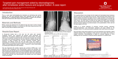
A 45-year-old female had a fall onto her left ankle with resultant trimalleolar fracture. She underwent left posterior ankle arthroscopy and debridement, and posterior malleolus open reduction internal fixation. At 2-week follow up, she reported increased foot pain and vasomotor changes. She returned at 6 weeks, with continued symptoms, and was referred to physical therapy. She continued to report increased pain, burning, violaceous changes, edema, weakness, and stiffness around her foot and ankle. Exam was notable for hyperesthesia in the lateral and anterior aspects of the dorsal foot, foot and ankle edema, and decreased ankle range of motion. X-rays at 2, 3, and 5 month follow up showed good healing with intact hardware.
She was diagnosed with complex regional pain syndrome (CRPS) type 1 and referred to pain management, who recommended a spinal cord stimulator trial. Near this time, she was also sent for an electromyography/nerve conduction study, which showed left superficial fibular and sural neuropathy, without other pathology. She underwent two positive sural nerve blocks with pain relief. She is planned to undergo left superficial nerve decompression with possible sural nerve targeted muscle reinnervation.
The superficial fibular nerve divides into the medial and intermediate dorsal cutaneous nerves of the foot at the distal third of the leg. These branches travel anterior to the lateral malleolus and divide into digital nerves to supply sensation to the dorsal foot, except for the first webspace (deep fibular nerve), and the lateral aspect of the fifth digit (sural nerve). The sural nerve is formed by the medial and lateral sural cutaneous nerves, which arise from the tibial and common fibular nerves, respectively. It courses behind the lateral malleolus to provide sensation to the posterolateral, inferior calf and the lateral aspect of the foot.
(CRPS) is a clinical diagnosis of exclusion involving sensory, vasomotor, sudomotor/edematous, and motor/trophic symptoms and signs detailed by the Budapest criteria. It is a well-documented complication after ankle fracture and surgery.
One must keep a high clinical suspicion for sural and superficial nerve damage in lateral malleolus fractures. Risk of damage to these nerves is even higher with a history of surgical intervention. Electrodiagnosis is helpful for accurate diagnosis of nerve pathology and can contribute to a tailored treatment approach for a patient's pain.
References: