425 posters, 1 videos, 9 audios, 8 topics, 53 sessions, 340 authors, 286 institutions
ePostersLive by SciGen Technologies S.A. All rights reserved.
22nd Annual Pain Medicine Meeting 2023
November 10 - 11, 2023 | New Orleans Marriott | New Orleans, Louisiana, USA

4733
PARAPLEGIA FOLLOWING AN INTRATHECAL HYDROMORPHONE AND BUPIVACAINE PUMP REMOVAL
Poster Presenter
Authors
Affiliations
Part of Topic
Medically Challenging Cases
Paraplegia Following an Intrathecal Hydromorphone and Bupivacaine Pump Removal
BACKGROUND: Intrathecal analgesia is a valuable treatment option for patients experiencing intractable pain that have failed more conservative management. Compared to other routes of drug delivery, intrathecal analgesia has been shown to have improved pain management with fewer systemic drug side effects.[1] In addition to pain relief, intrathecal pain management improves function and quality of life, with significant patient satisfaction.[2]
Here we present a rare case of incomplete paralysis in a patient during an intrathecal pain pump removal. This study emphasizes the significance of recognizing and managing the potential complications associated with intrathecal therapy for pain management.
CASE: A 36-year-old female with history of T12-S1 interbody fusion and chronic back pain managed with intrathecal hydromorphone and bupivacaine that had significantly reduced efficacy following a fall onto her back. Her pump was deemed fully functional via dye test and she subsequently was scheduled for extension of thoracolumbar fusion and pump replacement.
Intraoperatively, the pump catheter was found to be broken with an underlying collection of clear fluid, thought to be either medication or cerebrospinal fluid, which was subsequently removed by suction.
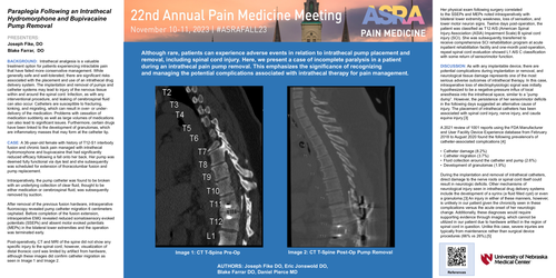
After removal of the previous fusion hardware, intraoperative fluoroscopy revealed that the pump catheter had migrated 6 centimeters cephalad. Before completion of the fusion extension, intraoperative electromyography revealed reduced somatosensory evoked potentials (SSEPs) and absent motor evoked potentials (MEPs) in the bilateral lower extremities and the operation was terminated early. Post-operatively, computed tomography and magnetic resonance imaging of the spine did not show any specific injury to the spinal cord, however, visualization of distal thoracic cord was limited by artifact from hardware, although these images did confirm catheter migration seen in Image 1 (pre-op) and Image 2 (post-op).
Her physical exam following surgery correlated to the SSEPs and MEPs noted intraoperatively with bilateral lower extremity weakness, loss of sensation, and lower motor neuron signs. Twelve days post-operation, the patient was classified as T12 AIS (American Spinal Injury Association (ASIA) Impairment Scale) B spinal cord injury (SCI). She was subsequently transferred to receive comprehensive SCI rehabilitation program at acute inpatient rehabilitation facility and one-month post-operation, repeat spinal cord evaluation showed L1 AIS C classification with some return of sensorimotor function.
DISCUSSION:
As with any implantable device, there are potential complications during implantation or removal, and neurological tissue damage represents one of the most serious adverse outcomes of intrathecal therapy. In this case, intraoperative loss of electrophysiologic signal was initially hypothesized to be a negative-pressure influx of local anesthesia into the intrathecal space, similar to a “pump dump”. However, the persistence of her sensorimotor deficits in the following days suggested an alternative cause of injury. The placement of intrathecal catheters has been associated with spinal cord injury, nerve injury, and cauda equina injury.[3]
A2021 review of 1001 reports using the FDA Manufacturer and User Facility Device Experience database from February 2018 to August 2020 found the following prevalence's of catheter-associated complications [4]:
During the implantation and removal of intrathecal catheters, direct damage to the nerve roots or spinal cord itself could result in neurologic deficits. Other mechanisms of neurological injury seen in intrathecal drug delivery systems include the development of a syrinx (a fluid filled cyst) or even a granuloma.[3] An injury in either of these manners, however, is unlikely in our patient given the chronicity seen in these complications versus the acute onset of her neurologic change. Additionally, these diagnoses would require supporting evidence through imaging, which cannot be utilized for in our patient due to hardware artifact in the region of spinal cord in question. Unlike this case, severe injuries are typically from maintenance rather than surgical device procedures (66% vs 26%).[5]