425 posters, 1 videos, 9 audios, 8 topics, 53 sessions, 340 authors, 286 institutions
ePostersLive by SciGen Technologies S.A. All rights reserved.
22nd Annual Pain Medicine Meeting 2023
November 10 - 11, 2023 | New Orleans Marriott | New Orleans, Louisiana, USA

4584
SACROILIAC JOINT RADIOFREQUENCY ABLATION POST SACROILIAC JOINT FUSION
Poster Presenter
Authors
Affiliations
Part of Topic
Medically Challenging Cases
Sacroiliac Joint Radiofrequency Ablation Post Sacroiliac Joint Fusion
Introduction
Sacroiliac joint dysfunction (SIJD) contributes to around 25% of reported cases of lower back discomfort in the general population, and this figure increases to approximately 37% in individuals who have previously undergone lumbosacral fusion surgery. Various factors can trigger sacroiliac joint (SI) pain, including trauma, pregnancy, stress, history of lumbar fusion surgery, and the use of bone grafts sourced from areas near the SI. The presentation of SIJD symptoms can often resemble other types of lower back pain, emphasizing the importance of accurate diagnosis and effective treatment. A comprehensive assessment relies heavily on the patient’s history and thorough physical examination.
Pain arising from the SI area typically manifests as discomfort below the beltline, with potential radiation towards the groin or lower extremities. In some cases, patients might report pain radiating below the knee within the L5-S1 dermatome region. Additionally, common symptoms cited by patients include pain during sitting, lying on the affected side, or prolonged walking. To diagnose SIJD, various provocative tests such as the thigh thrust test, FABER test, compression test, Gaenslen test, Sacral thrust test, distraction test, and the Fortin finger test are employed.
A definitive gold standard physical examination for SIJD remains elusive. Research demonstrates that a combination of provocative tests yields superior results compared to individual tests. Positive outcomes are more prevalent when three or more tests are employed. Notably, the combination of FABER and thigh thrust tests demonstrates the highest accuracy, closely followed by the combination of FABER and Gaenslen's test. In terms of imaging techniques, MRI is the most sensitive method for diagnosing SIJD, capable of revealing bone marrow edema and inflammation surrounding the joint.
The key diagnostic confirmation for SIJD is the SI joint diagnostic block. A positive indication for the source of SI joint pain is considered when this procedure results in a 50% to 75% reduction of pain.
Case Report
A 67-year-old male with a medical history that includes Sarcoidosis, DVT on Xarelto, Prostate cancer, and chronic low back pain was referred to our clinic for an assessment of persistent pain in the right back and hip. He had undergone several surgeries in the past, including L4-S1 foraminotomy, L3-S1 instrumentation, and bilateral open SI joint arthrodesis. Despite some initial relief following these surgeries, his pain progressively worsened over time.
Upon examination, the patient reported experiencing varying levels of pain in his right gluteal region, particularly aggravated when lying down or sitting. He had previously attempted physical therapy and continued home exercises, as well as using an SI belt, which provided partial relief. His current medication regimen included Tramadol, Gabapentin, Baclofen, Flexeril, and Lidocaine patches. Previous interventions consisted of a right hip bursa injection and a right SI joint injection, resulting in partial relief.
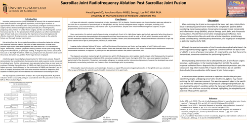
Imaging studies indicated bilateral SI fusion, multilevel lumbosacral laminectomy and fusion, and narrowing of both SI joints with more pronounced sclerosis on the right side. Limited osseous fusion was observed along the superior right SI joint. Considering the inadequate response to conservative treatments, a diagnostic SI joint injection was planned to address the patient's persistent pain.
The diagnostic procedure involved a right SI joint injection with 0.25% Bupivacaine, which yielded significant pain relief. Subsequently, the patient was scheduled for a Radiofrequency Ablation (RFA) procedure to ablate the right SI joint. During the RFA, three sites were targeted, resulting in partial relief of the discomfort. The patient expressed a willingness to undergo another interventional procedure; however, he developed new-onset bradycardia, necessitating evaluation and clearance from his cardiologist prior to proceeding.
Following the required evaluation and cardiologist clearance, a repeat RFA procedure targeting three sites in the right SI joint was scheduled. After a span of 14 days, the patient reported notable improvement in his pain levels.
Discussion
After confirming the SI joint as the origin of the lower back pain, initial efforts focus on employing conservative treatments for symptomatic patients before considering more invasive options. Conservative measures include nonsteroidal anti-inflammatory drugs (NSAIDs), physical therapy, pelvic belts, and chiropractic manipulations. Should these conservative strategies prove ineffective, more advanced interventions like extra and intra-articular injections, prolotherapy, pulsed radiofrequency, radiofrequency denervation, and surgery can be explored as potential treatments for SIJD.
Although the precise innervation of the SI remains incompletely elucidated, the prevailing understanding suggests a significant contribution from the dorsal rami of L4 to S3 in terms of sensory innervation. It's important to note that there isn't a standardized procedure established for performing SI joint RFA.
When preceding interventions fail to alleviate the pain, SI joint fusion surgery becomes a viable option. In the treatment algorithm for SIJD, if a positive response is obtained from a diagnostic block in patients with persistent pain, the subsequent step often involves RFA. If repeat interventions do not yield the desired relief, the consideration shifts toward SI joint fusion surgery.
In situations where patients continue to experience intolerable pain post-operatively despite undergoing conservative treatments, options may include revisiting the SIJD treatment protocol. This was the approach taken in the specific case mentioned, where the patient had previously undergone SI joint fusion. By reevaluating the patient's condition within the framework of the SIJD treatment algorithm, pain relief was successfully achieved, highlighting the adaptability and potential efficacy of this approach.
References
Aydin, S.M. et al. (2010) ‘The role of radiofrequency ablation for sacroiliac joint pain: A meta-analysis’, PM&R, 2(9), pp. 842–851. doi:10.1016/j.pmrj.2010.03.035.
Buchanan, P. et al. (2021) ‘Successful diagnosis of sacroiliac joint dysfunction’, Journal of Pain Research, Volume 14, pp. 3135–3143. doi:10.2147/jpr.s327351.
Falowski, S. et al. (2020) ‘<P>A review and algorithm in the diagnosis and treatment of sacroiliac joint pain</p>’, Journal of Pain Research, Volume 13, pp. 3337–3348. doi:10.2147/jpr.s279390.
Lee, A. et al. (2022) ‘Sacroiliitis: A review on anatomy, diagnosis, and treatment’, Advances in Orthopedics, 2022, pp. 1–8. doi:10.1155/2022/3283296.