425 posters, 1 videos, 9 audios, 8 topics, 53 sessions, 340 authors, 286 institutions
ePostersLive by SciGen Technologies S.A. All rights reserved.
22nd Annual Pain Medicine Meeting 2023
November 10 - 11, 2023 | New Orleans Marriott | New Orleans, Louisiana, USA

4592
MALARIA INDUCED GLOSSOPHARYNGEAL NEURALGIA MANAGED WITH ROUTINE NERVE BLOCKS
Poster Presenter
Authors
Affiliations
Part of Topic
Medically Challenging Cases
Atypical Glossopharyngeal Neuralgia Managed with Nerve Blocks FollowingMefloquine Treatment of Malaria
A 38 year old healthy female developed bilateral glossopharyngeal neuralgia with hemiplegic migraines after being treated with mefloquine for a severe P. falciparum infection (4/2016) while living in Africa working with gorillas and primates. Symptoms were described as severe “electric-sharp-stabbing pain” (rated pain 8-9/10) and included facial spasms, trouble speaking/raspy voice, dysphagia and facial weakness. Symptoms initially responded to a course of steroids, but eventually persisted to the point where she developed weight loss due to aversion to food and the inability to perform activities of daily living.
Initial management:
Botox injections, physical therapy, multimodal medications (duloxetine, gabapentin, acetaminophen, NSAIDS), bilateral cervical facet injections, occipital nerve blocks, which provided ~50% pain relief for up to 1-2 weeks. However, painful throat spasms persisted, and her symptoms became refractory to initial treatment.
Bilateral Glossopharyngeal Nerve Blocks and Stellate Ganglion Blocks
-Provided effective relief reported around 50 %, pain returning to baseline level by 3 months. Repeat injections reduced level of pain from 9-10 (2021) to 3-4 in (2023).
-Patient regained the ability to eat and drink, no longer had facial muscle spasms, significant improvement in overall quality of life
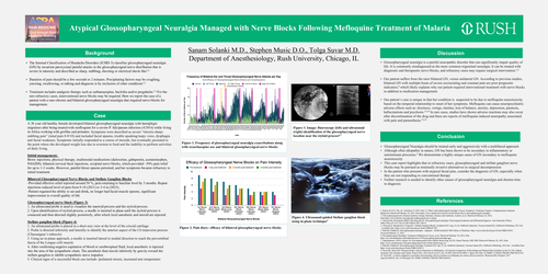
Glossopharyngeal nerve block (Figure 3)
1. An ultrasound probe is used to visualize the mastoid process and the styloid process.
2. Upon identification of styloid process, a needle is inserted in plane until the styloid process is contacted and then directed slightly posteriorly, after which local anesthetic and steroid are injected
Stellate ganglion block (Figure 4)
1. An ultrasound probe is placed in a short axis view at the level of the cricoid cartilage
2. Probe is directed inferiorly and laterally to identify the anterior aspect of the C6 transverse process (Chassaignac’s tubercle)
3. Using an in-plane approach, a needle is inserted lateral to medial direction to reach the prevertebral fascia of the Longus colli muscle
4. After confirming negative aspiration of blood or cerebrospinal fluid, local anesthetic is injected into the area of the sympathetic chain. The anesthetic then travels inferiorly by gravity toward the stellate ganglion to inhibit sympathetic nerve impulses
5. Clinical signs of a successful block can include: ipsilateral miosis, increased arm temperature