425 posters, 1 videos, 9 audios, 8 topics, 53 sessions, 340 authors, 286 institutions
ePostersLive by SciGen Technologies S.A. All rights reserved.
22nd Annual Pain Medicine Meeting 2023
November 10 - 11, 2023 | New Orleans Marriott | New Orleans, Louisiana, USA

4693
SPONTANEOUS SPINAL EPIDURAL HEMATOMA FOLLOWING HEAVY WEIGHTLIFTING: A CASE REPORT
Poster Presenter
Authors
Affiliations
Part of Topic
Medically Challenging Cases
Spontaneous Spinal Epidural Hematoma Following Heavy Weightlifting: A Case Report
Introduction: Spontaneous spinal epidural hematoma (SSEH) refers to extradural hemorrhage without a known precipitating cause such as trauma, tumor, or vascular malformation. The incidence of SSEH has been estimated at 0.1 in 100,000 individuals per year. SSEH acutely presents as severe neck or back pain with dermatomal radiation to the extremities. It may cause sensory changes, paresis, and bowel or bladder dysfunction if the spinal cord or cauda equina is compressed. Valsalva maneuvers during weightlifting increase intraabdominal and intrathoracic pressures which are ultimately transferred to the spinal epidural venous system. This increase in pressure has been attributed to rupture and hemorrhage of the epidural venous vessels resulting in SSEH.
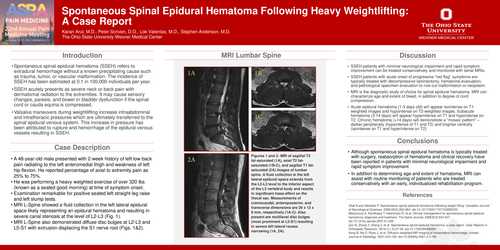
Case Description: A 48-year-old male presented with 2-week history of left low back pain radiating to the left anteromedial thigh and weakness of left hip flexion. He reported percentage of axial to extremity pain as 25% to 75%. He was performing a heavy weighted exercise of over 320 lbs (known as a seated good morning) at time of symptom onset. Examination remarkable for positive seated left straight leg raise and left slump tests. MRI L-Spine showed a fluid collection in the left lateral epidural space likely representing an epidural hematoma and resulting in severe canal stenosis at the level of L2-L3 (Fig. 1). MRI L-Spine also demonstrated diffuse disc bulges at L2-L3 and L5-S1 with extrusion displacing the S1 nerve root (Figs. 1&2).
Figures 1 and 2: MRI of sagittal T2 fat-saturated (1A), axial T2 fat-saturated (1B-C), and sagittal T1 fat-saturated (2A) images of lumbar spine. A fluid collection in the left lateral epidural space extends from the L2-L3 level to the inferior aspect of the L3 vertebral body and results in significant mass effect on the thecal sac. Measurements of craniocaudal, anteroposterior, and transverse dimensions are 29 x 12 x 9 mm, respectively (1A-C). Also present are multilevel disc bulges (most prominent at L5-S1) resulting in severe left lateral recess narrowing (1A, 2A).