66 posters, 2 videos, 1 audios, 2 topics, 162 authors, 49 institutions
ePostersLive by SciGen Technologies S.A. All rights reserved.
American College of Prosthodontists - 2020 ANNUAL SESSION
November 4-7 | Virtual Meeting

49
Observation of ion depletion region on the enamel during electrokinetic flow application.
Poster Presenter
Authors
Affiliations
Part of Topic
Original Research
Introductio
Direct infiltration of various molecules/ions into dental enamel is highly desirable in dentistry, but the nanometric scale of enamel pores inherently limits it3,4,6. Certain dental procedures such as dental bleaching and resin infiltration rely on diffusion and capillarity, and they often require relatively long application time to be effective. Our group demonstrated the use of electrokinetic flow (EKF) to deliver ions into the enamel to significantly greater depth compared to diffusion alone, reaching up to 1mm1,2. Electrokinetic flow (EKF), as demonstrated by Schoch et al., enables the movement of liquid throughnanoporous materials with the application of the electric field. According to Gan et al., electrokinetically enhanced direct resin infiltration to enamel was recorded up to 2,000 um. Unfortunately, it takes about 30 min for millimeter-scale infiltration1making it less practical, although it has enormous potential. The underlying multi-physics of the electrokineticphenomenon on enamel is still unclear, preventing optimization and further improvement merely because it is hard to observe. This study observed the ion depletion phenomenon around enamel during electrokinetic flow application using a microfluidic device. This evidence suggests that material transport using electrokinetic flow may significantly be affected by ions near the enamel surface during the EKF application.
Materials and Methods
Caries free tooth sample was selected and hydrated in formalin solution. The tooth sample was sectioned in a mesiodistal direction using a diamond disk. The tooth section was carefully examined under a microscope to confirm the absence of any cracks. The sectioned sample was then polished with sand-papers in 400, 600, 800, and 1200 grit.
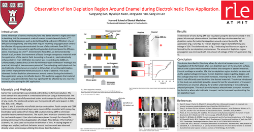
Figure 1. and 2. show the microfluidic device construction. Tooth sample and CEM (cation exchange membrane) layer were mounted then insulated with epoxy clay. Electrodes were isolated from Alexa 488 solution utilizing CEM to eliminate possible electrochemical reactions. The acrylic layer with four channels was added for mechanical support. Four electrodes were placed through the channel for probing electric current and application of voltage. Alex 488 dye (ThermoFisher Scientific, inc.) was used to visualize the behavior of ions. A varying degree of voltage was applied, and ionic behavior around tooth enamel was observed directly under a microscope utilizing the device described above.
Results
The behavior of ions during EKF was visualized using the device described in this paper. Microscopic observation of the Alexa 488 dye solution revealed ion depletion region formation/propagation around enamel during the EKF application (Fig. 3 and Fig. 4). The ion depletion region started forming at a voltage of 10V. The darkened area in fig. 3 indicating less fluorescent signal is formed by the ion depletion phenomenon. The amount of depletion region propagation was increased with the amount of voltage of the EKF application (fig. 4).
Concusion
The device described in this study allows for electrical measurement and visualization of the formation of an ion-depletion layer on the enamel’s surface, which is the culprit mechanism that hinders the transport of desired materials. Even at a voltage as small as 10V, the ion-depletion region starts to be observed. As the applied voltage increases, the ion depletion region is getting bigger, and thus voltage drop near the enamel increases, meaning that most of the electric field is not efficiently used to deliver the desired materials. The device introduced in this study can potentially provide a powerful tool for visualization of transport phenomena on enamel, allowing for a better understanding of the underlying physical principles. This result directly impacts electrokinetic transport research for dentistry, where electrokinetic transport can be improved by minimizing the ion-depletion layer.
References