1,267 posters, 47 videos, 13 topics, 4 sessions, 853 authors
ePostersLive by SciGen Technologies S.A. All rights reserved.
SOHO 2026 Annual Meeting
September 9 - 12, 2026 | George R. Brown Convention Center, Houston, Texas

CLL - 1564
Concurrent Hemophagocytosis and Hodgkin Lymphoma-Type Richter Transformation in CLL/SLL: An Incidental Finding With Therapeutic Implications
Poster Presenter
Authors
Part of Topic
Chronic Lymphocytic Leukemia (CLL)
KEYWORDS: Chronic lymphocytic leukemia, Richter transformation, Hodgkin lymphoma, hemophagocytic lymphohistiocytosis, bone marrow biopsy
INTRODUCTION: Richter transformation (RT) to classic Hodgkin lymphoma (CHL) accounts for ~10% of RT cases in chronic lymphocytic leukemia/small lymphocytic leukemia (CLL/SLL). Hemophagocytic lymphohistiocytosis (HLH) complicating CLL/SLL is exceedingly rare, with fewer than five reported cases. We report the first case of incidental hemophagocytosis discovered during bone marrow workup for suspected RT in CLL/SLL, subsequently confirmed by HLH biomarkers.
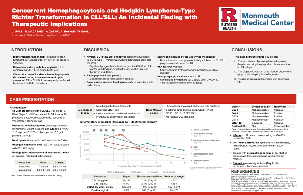
CASE PRESENTATION: An 85-year-old female with CLL/SLL with progression on Acalabrutinib, and is currently on Venetoclax + Obinituzumab presented with B-symptoms and pancytopenia. Mild splenomegaly and interval reduction of mediastinal lymphadenopathy from previous were seen on imaging. Despite radiographic improvement, high clinical suspicion for RT prompted lymph node biopsy. This yielded non-diagnostic tissue fragments with equivocal EBER-ISH; lesional cells were lost on deeper sections precluding PAX5/CD30 confirmation.
Subsequent bone marrow biopsy revealed hypercellular marrow with grade 2 reticulin fibrosis and scattered large cells (CD20−, PAX5+, CD30+, CD15−, EBER-ISH−) suggestive of CHL-type RT, with no residual CLL. Incidental histiophagocytosis was identified, with elevated sCD25 >4000 U/mL, CXCL9 10,159 pg/mL, and IL-18 6,847 pg/mL. HLH-94 protocol was initiated with dexamethasone 10 mg/m²; rising inflammatory markers necessitated age-adjusted etoposide (50 mg/m²).
DISCUSSION: This case represents a previously unreported triad of suspected CD15-negative HL-type RT, non-diagnostic lymph node biopsy requiring bone marrow-based diagnosis, and incidental hemophagocytosis. The atypical CD15−/EBER− phenotype raises the question of true CHL-type RT versus CLL with Hodgkin/Reed-Sternberg-like cells, a distinction with significant prognostic implications (median OS 57 vs. 8.4 months with Hodgkin-directed therapy). The paradox of radiographically improving lymphadenopathy despite clinical deterioration further underscores the limitations of imaging alone in RT surveillance. Constitutional symptoms and cytopenias were attributed to CLL/SLL progression and suspected RT, highlighting the diagnostic challenge when HLH features overlap with the underlying malignancy. Specialized biomarkers (sCD25, CXCL9, IL-18) provided critical confirmatory evidence and may distinguish true HLH from malignancy-mimicking features in CLL/SLL.
CONCLUSION: This case highlights three key points: (1) the importance of pursuing tissue diagnosis despite improving imaging when clinical suspicion for RT is high; (2) the diagnostic value of bone marrow biopsy when lymph node sampling is nondiagnostic; and (3) the role of specialized biomarkers in confirming HLH.