816 posters, 24 topics, 3,314 authors, 1,012 institutions
ePostersLive by SciGen Technologies S.A. All rights reserved.
SAGES 2026 Annual Meeting Emerging Technology Session
March 25-28, 2026 | Tampa, FL, USA

ETP897
From Scan to Scope: Translating Imaging into Intraoperative Insight
Poster Presenter
Authors
Affiliations
Part of Topic
From Scan to Scope: Translating Imaging into Intraoperative Insight
Delorme, Sebastien, PhD1; Ahmed, Hasnaien, MD2; Noreau, Antoine, P.Eng.3
1. Scopia Surgical Inc., Montreal, Canada
2. University of Alberta, Edmonton, Canada
3. Scopia Surgical Inc., Montreal, Canada
Contact email: antoine@scopia.tech
Context
Anatomic variability and complex tumor relationships present significant challenges for both surgical planning and execution. An accurate interpretation of preoperative imaging by the surgeon is crucial for better oncological outcomes [1]. Current practice often relies on 3D-printed [2] or mental reconstruction of 3D anatomy, the latter of which increases cognitive demand and uncertainty during surgery.
Objective
This project focuses on low anterior resections in colorectal surgery and aims at generating:
-
patient-specific 3D digital models for:
-
preoperative planning
-
intraoperative reference.
Anticipated benefits include:
-
surgical efficiency,
-
improved circumferential resection margins (CRM),
From Scan to 3D Model
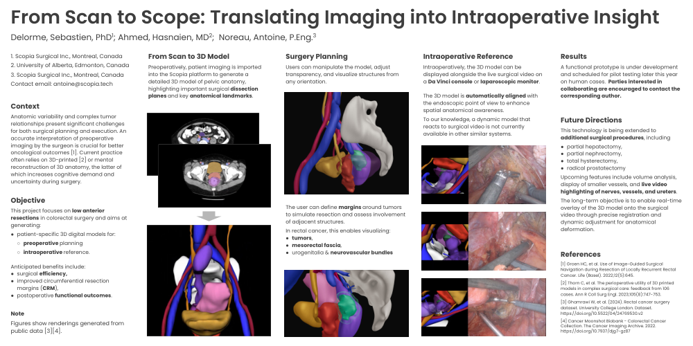
Preoperatively, patient imaging is imported into the Scopia platform to generate a detailed 3D model of pelvic anatomy, highlighting important surgical dissection planes and key anatomical landmarks.
Surgery Planning
Users can manipulate the model, adjust transparency, and visualize structures from any orientation.
The user can define margins around tumors to simulate resection and assess involvement of adjacent structures.
In rectal cancer, this enables visualizing:
-
tumors,
-
mesorectal fascia,
-
urogenitalia & neurovascular bundles
Intraoperative Reference
Intraoperatively, the 3D model can be displayed alongside the live surgical video on a Da Vinci console or laparoscopic monitor.
The 3D model is automatically aligned with the endoscopic point of view to enhance spatial anatomical awareness.
To our knowledge, a dynamic model that reacts to surgical video is not currently available in other similar systems.
Results
A functional prototype is under development and scheduled for pilot testing later this year on human cases. Parties interested in collaborating are encouraged to contact the corresponding author.
Future Directions
This technology is being extended to additional surgical procedures, including
-
partial hepatectomy,
-
partial nephrectomy,
-
total hysterectomy,
-
radical prostatectomy
Upcoming features include volume analysis, display of smaller vessels, and live video highlighting of nerves, vessels, and ureters.
The long-term objective is to enable real-time overlay of the 3D model onto the surgical video through precise registration and dynamic adjustment for anatomical deformation.
References
[1] Groen HC, et al. Use of Image-Guided Surgical Navigation during Resection of Locally Recurrent Rectal Cancer. Life (Basel). 2022;12(5):645.
[2] Thorn C, et al. The perioperative utility of 3D printed models in complex surgical care: feedback from 106 cases. Ann R Coll Surg Engl. 2023;105(8):747-753.
[3] Ghamrawi W, et al. (2024). Rectal cancer surgery dataset. University College London. Dataset. https://doi.org/10.5522/04/24769530.v2
[4] Cancer Moonshot Biobank - Colorectal Cancer Collection. The Cancer Imaging Archive. 2022. https://doi.org/10.7937/djg7-gz87