816 posters, 24 topics, 3,314 authors, 1,012 institutions
ePostersLive by SciGen Technologies S.A. All rights reserved.
SAGES 2026 Annual Meeting Emerging Technology Session
March 25-28, 2026 | Tampa, FL, USA

ETP875
NeuVue Perfusion: Geometry-Corrected Quantitative Fluorescence Mapping for Real-Time Assessment of Tissue Viability
Part of Topic
NeuVue Perfusion: Geometry-Corrected Quantitative Fluorescence Mapping for Real-Time Assessment of Tissue Viability
Objective:
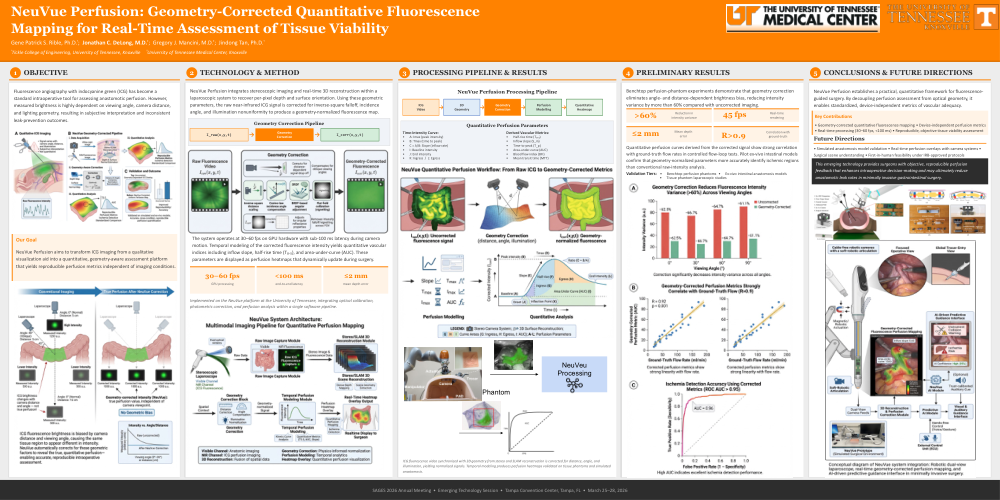
Fluorescence angiography with indocyanine green (ICG) has become a standard intraoperative tool for assessing anastomotic perfusion. However, measured brightness is highly dependent on viewing angle, camera distance, and lighting geometry, resulting in subjective interpretation and inconsistent leak-prevention outcomes. NeuVue Perfusion aims to transform ICG imaging from a qualitative visualization aid into a quantitative, geometry-aware assessment platform that yields reproducible perfusion metrics independent of imaging conditions.
Description of Technology and Method:
NeuVue Perfusion integrates stereoscopic imaging and real-time 3D reconstruction within a laparoscopic system to recover per-pixel depth and surface orientation. Using these geometric parameters, the raw near-infrared ICG signal is corrected for inverse-square falloff, incidence angle, and illumination nonuniformity to produce a geometry-normalized fluorescence map. The system operates at 30–60 frames per second on GPU hardware, maintaining sub-100 ms latency during camera motion. Temporal modeling of the corrected fluorescence intensity yields quantitative vascular indices including inflow slope, half-rise time, and area-under-curve (AUC). These parameters are displayed as perfusion heatmaps that dynamically update during surgery. The method is implemented on the NeuVue platform developed at the University of Tennessee, integrating optical calibration, photometric correction, and perfusion analysis within a single software pipeline.
Preliminary Results:
Benchtop perfusion-phantom experiments demonstrate that geometry correction eliminates angle- and distance-dependent brightness bias, reducing intensity variance by more than 60 % compared with uncorrected imaging. Real-time rendering of corrected fluorescence achieves 45 fps with mean depth error ≤ 2 mm. Quantitative perfusion curves derived from the corrected signal show strong correlation (R > 0.9) with ground-truth flow rates in controlled flow-loop tests. Pilot ex-vivo intestinal models confirm that geometry-normalized parameters more accurately identify ischemic regions than conventional raw-intensity analysis.
Conclusions / Future Directions:
NeuVue Perfusion establishes a practical, quantitative framework for fluorescence-guided surgery. By decoupling perfusion assessment from optical geometry, it enables standardized, device-independent metrics of vascular adequacy. Ongoing work is extending validation to simulated laparoscopic anastomosis models and integrating real-time perfusion overlays with robotic camera systems. This emerging technology provides surgeons with objective, reproducible perfusion feedback that enhances intraoperative decision-making and may ultimately reduce anastomotic leak rates in minimally invasive gastrointestinal surgery.