875 posters, 25 topics, 3,440 authors, 1,061 institutions
ePostersLive by SciGen Technologies S.A. All rights reserved.
SAGES 2026 Annual Meeting Emerging Technology Session
March 25-28, 2026 | Tampa, FL, USA

P104
Preventing Unwarranted Diagnostic Cascades: Lessons from Central Venous Catheter Placement in a Patient with Arteriovenous Fistula
Poster Presenter
Part of Topic
Acute Care
Ipsilateral Arteriovenous Fistula Outflow Mimicking Inadvertent Carotid Artery Puncture: A Diagnostic Dilemma in Central Venous Access
Austin Chao, BA1; Enrico Camporesi, MD{1,2}; Elia Elia, MD2
1USF Health Morsani College of Medicine, Tampa, FL
2Department of Anesthesiology and Perioperative Medicine, Tampa General Hospital, Tampa, FL
Objective: To demonstrate how placing a central venous catheter (CVC) ipsilateral to a high-flow arteriovenous fistula (AVF) causes expected elevations in central venous pressure (CVP) and oxygen saturation (ScvO2), guiding a rational diagnostic workup and preventing the immediate misdiagnosis of arterial cannulation.
Introduction: The right internal jugular (IJ) vein is the preferred site for CVC placement due to its linear trajectory to the cavoatrial junction. When right-sided access is unavailable, the left IJ is utilized, requiring the catheter to navigate the left brachiocephalic vein across the midline. This tortuous path increases the risk of malposition and stenosis. In patients with an existing AVF, CVCs should ideally be placed contralateral to the access arm. A high-flow AVF shunts high-pressure, highly oxygenated arterial blood directly into the central venous system. Placing a CVC ipsilateral to an AVF causes the catheter to sample this arterialized venous return, particularly when the tip terminates shallowly before the superior vena cava. This localized physiologic alteration mimics the exact presentation of an inadvertent arterial cannulation by producing markedly elevated CVP and an abnormally high ScvO2 (>80%). Recognizing this expected hemodynamic and biochemical shift is critical for the anesthesia and surgical teams to guide bedside triage and prevent unnecessary emergent surgical interventions for suspected arterial injury.
Case Presentation
Patient: A 57-year-old male with end-stage renal disease on hemodialysis via a left upper extremity (LUE) AVF presented for a deceased donor kidney transplant.
Intraoperative Course: Prior to incision, a 7-French triple-lumen CVC was placed in the left IJ vein under ultrasound guidance by the anesthesia team. Intravascular placement was confirmed via ultrasound and pressure transduction. The transplant proceeded without intraoperative complication.
Complication: In the PACU, the patient became hypotensive (MAPs in the 80s) with right lower quadrant tenderness and 150 cc of sanguinous drain output. This prompted aggressive resuscitation for a suspected surgical hematoma.
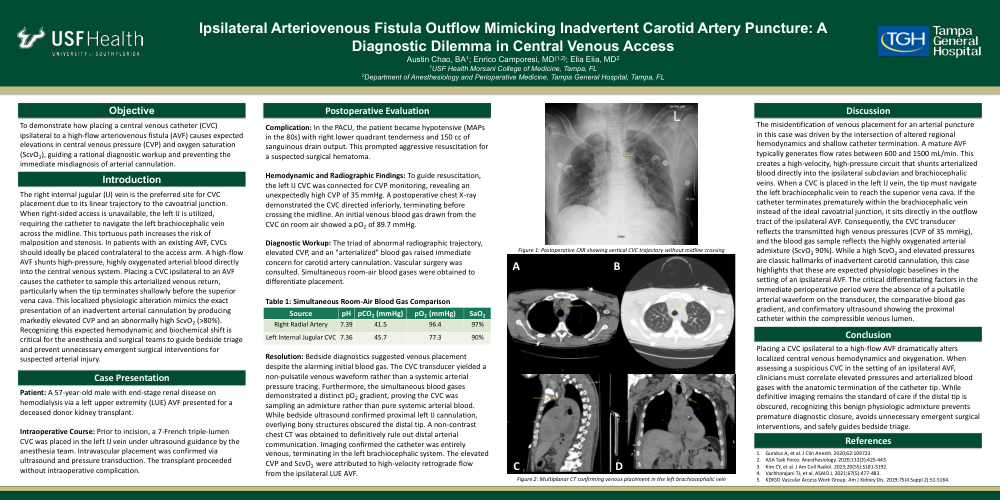
Hemodynamic and Radiographic Findings: To guide resuscitation, the left IJ CVC was connected for CVP monitoring, revealing an unexpectedly high CVP of 35 mmHg. A postoperative chest X-ray demonstrated the CVC directed inferiorly, terminating before crossing the midline. An initial venous blood gas drawn from the CVC on room air showed a pO2 of 89.7 mmHg.
Diagnostic Workup: The triad of abnormal radiographic trajectory, elevated CVP, and an "arterialized" blood gas raised immediate concern for carotid artery cannulation. Vascular surgery was consulted. Simultaneous room-air blood gases were obtained to differentiate placement.
|
Source |
pH |
pCO2 (mmHg) |
pO2 (mmHg) |
SaO2 |
|
Right Radial Artery |
7.39 |
41.5 |
96.4 |
97% |
|
Left Internal Jugular CVC |
7.36 |
45.7 |
77.3 |
90% |
Resolution: Bedside diagnostics suggested venous placement despite the alarming initial blood gas. The CVC transducer yielded a non-pulsatile venous waveform rather than a systemic arterial pressure tracing. Furthermore, the simultaneous blood gases demonstrated a distinct pO2 gradient, proving the CVC was sampling an admixture rather than pure systemic arterial blood. While bedside ultrasound confirmed proximal left IJ cannulation, overlying bony structures obscured the distal tip. A non-contrast chest CT was obtained to definitively rule out distal arterial communication. Imaging confirmed the catheter was entirely venous, terminating in the left brachiocephalic system. The elevated CVP and ScvO2 were attributed to high-velocity retrograde flow from the ipsilateral LUE AVF.
Discussion: The misidentification of venous placement for an arterial puncture in this case was driven by the intersection of altered regional hemodynamics and shallow catheter termination. A mature AVF typically generates flow rates between 600 and 1500 mL/min. This creates a high-velocity, high-pressure circuit that shunts arterialized blood directly into the ipsilateral subclavian and brachiocephalic veins. When a CVC is placed in the left IJ vein, the tip must navigate the left brachiocephalic vein to reach the superior vena cava. If the catheter terminates prematurely within the brachiocephalic vein instead of the ideal cavoatrial junction, it sits directly in the outflow tract of the ipsilateral AVF. Consequently, the CVC transducer reflects the transmitted high venous pressures (CVP of 35 mmHg), and the blood gas sample reflects the highly oxygenated arterial admixture (ScvO2 90%). While a high ScvO2 and elevated pressures are classic hallmarks of inadvertent carotid cannulation, this case highlights that these are expected physiologic baselines in the setting of an ipsilateral AVF. The critical differentiating factors in the immediate perioperative period were the absence of a pulsatile arterial waveform on the transducer, the comparative blood gas gradient, and confirmatory ultrasound showing the proximal catheter within the compressible venous lumen.
Conclusion: Placing a CVC ipsilateral to a high-flow AVF dramatically alters localized central venous hemodynamics and oxygenation. When assessing a suspicious CVC in the setting of an ipsilateral AVF, clinicians must correlate elevated pressures and arterialized blood gases with the anatomic termination of the catheter tip. While definitive imaging remains the standard of care if the distal tip is obscured, recognizing this benign physiologic admixture prevents premature diagnostic closure, avoids unnecessary emergent surgical interventions, and safely guides bedside triage.