119 posters, 6 topics, 524 authors, 243 institutions
ePostersLive by SciGen Technologies S.A. All rights reserved.
RCR Global AI Conference 2026
29-30 June, 2026 | QEII Centre, Westminster

111
Benchmarking CNNs and Transformers for Knee Cartilage MRI Segmentation Without Pre-Training
Poster Presenter
Authors
Affiliations
Part of Topic
AI Education and research: examples of proof of concept or AI in development, technical advances, teaching approaches or pre-clinical testing
Benchmarking CNNs and Transformers for Knee Cartilage MRI Segmentation Without Pre-Training
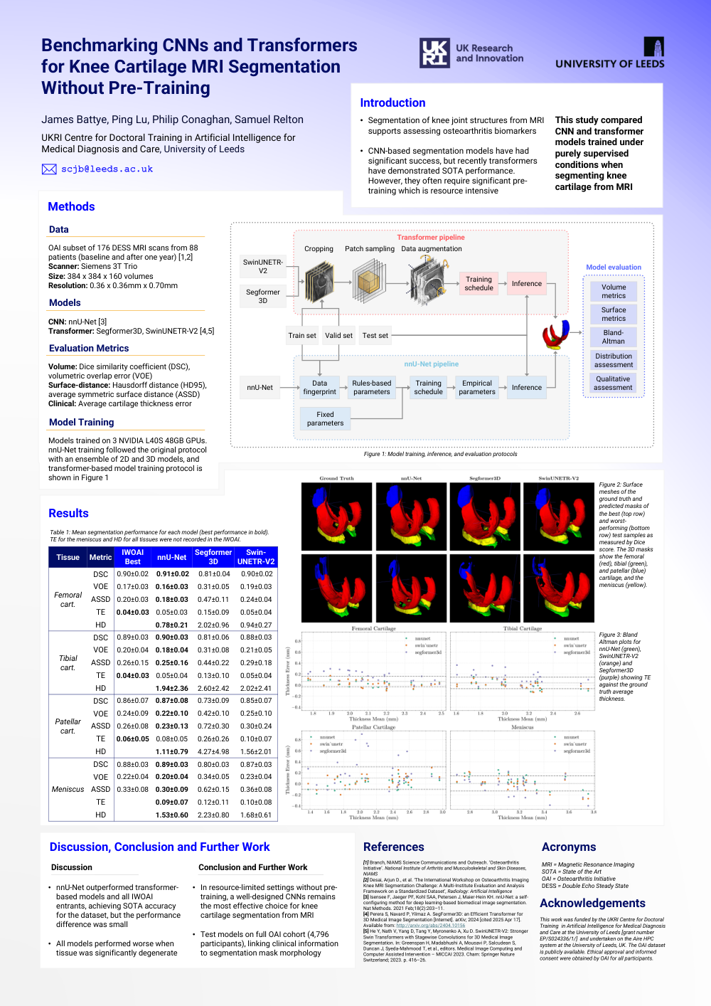
Data
OAI subset of 176 DESS MRI scans from 88 patients (baseline and after one year) [1,2]
Scanner: Siemens 3T Trio Size: 384 x 384 x 160 volumes
Resolution: 0.36 x 0.36mm x 0.70mm
Models
CNN: nnU-Net [3]
Transformer: Segformer3D, SwinUNETR-V2 [4,5]
Evaluation Metrics
Volume: Dice similarity coefficient (DSC), volumetric overlap error (VOE)
Surface-distance: Hausdorff distance (HD95), average symmetric surface distance (ASSD)
Clinical: Average cartilage thickness error
Model Training
Training schedule Empirical parameters Fixed parameters Models trained on 3 NVIDIA L40S 48GB GPUs. nnU-Net training followed the original protocol with an ensemble of 2D and 3D models, and transformer-based model training protocol is shown in Figure 1.
Discussion, Conclusion and Further Work
• nnU-Net outperformed transformer based models and all IWOAI entrants, achieving SOTA accuracy for the dataset, but the performance difference was small
• In resource-limited settings without pre training, a well-designed CNNs remains the most effective choice for knee cartilage segmentation from MRI
• All models performed worse when tissue was significantly degenerate
• Test models on full OAI cohort (4,796 participants), linking clinical information to segmentation mask morphology
Acronyms
MRI = Magnetic Resonance Imaging
SOTA = State of the Art
OAI = Osteoarthritis Initiative
DESS = Double Echo Steady State
Acknowledgements
This work was funded by the UKRI Centre for Doctoral Training in Artificial Intelligence for Medical Diagnosis and Care at the University of Leeds [grant number EP/S024336/1/] and undertaken on the Aire HPC system at the University of Leeds, UK. The OAI dataset is publicly available. Ethical approval and informed consent were obtained by OAI for all participants.
References
[1] Branch, NIAMS Science Communications and Outreach. ‘Osteoarthritis Initiative’. National Institute of Arthritis and Musculoskeletal and Skin Diseases, NIAMS [2] Desai, Arjun D., et al. ‘The International Workshop on Osteoarthritis Imaging Knee MRI Segmentation Challenge: A Multi-Institute Evaluation and Analysis Framework on a Standardized Dataset’, Radiology: Artificial Intelligence
[3] Isensee F, Jaeger PF, Kohl SAA, Petersen J, Maier-Hein KH. nnU-Net: a self configuring method for deep learning-based biomedical image segmentation. Nat Methods. 2021 Feb;18(2):203–11.
[4] Perera S, Navard P, Yilmaz A. SegFormer3D: an Efficient Transformer for 3D Medical Image Segmentation [Internet]. arXiv; 2024 [cited 2025 Apr 17]. Available from: http://arxiv.org/abs/2404.10156
[5] He Y, Nath V, Yang D, Tang Y, Myronenko A, Xu D. SwinUNETR-V2: Stronger Swin Transformers with Stagewise Convolutions for 3D Medical Image Segmentation. In: Greenspan H, Madabhushi A, Mousavi P, Salcudean S, Duncan J, Syeda-Mahmood T, et al., editors. Medical Image Computing and Computer Assisted Intervention – MICCAI 2023. Cham: Springer Nature Switzerland; 2023. p. 416–26