2,281 posters, 63 videos, 56 audios, 43 topics, 18 sessions, 6,174 authors, 2,323 institutions
ePostersLive by SciGen Technologies S.A. All rights reserved.
RCOG World Congress 2025
June 23 - 25, 2025 | ExCeL London, UK
EP.0051
Pregnancy and parvovirus B19 infection
Part of Topic
Advances in prenatal diagnosis
Introduction
Parvovirus B19 infection during pregnancy is a well-known cause of fetal hydrops, severe anemia and fetal death. Diagnosis and management require a careful and timely approach, involving both maternal and fetal monitoring.
Case report :
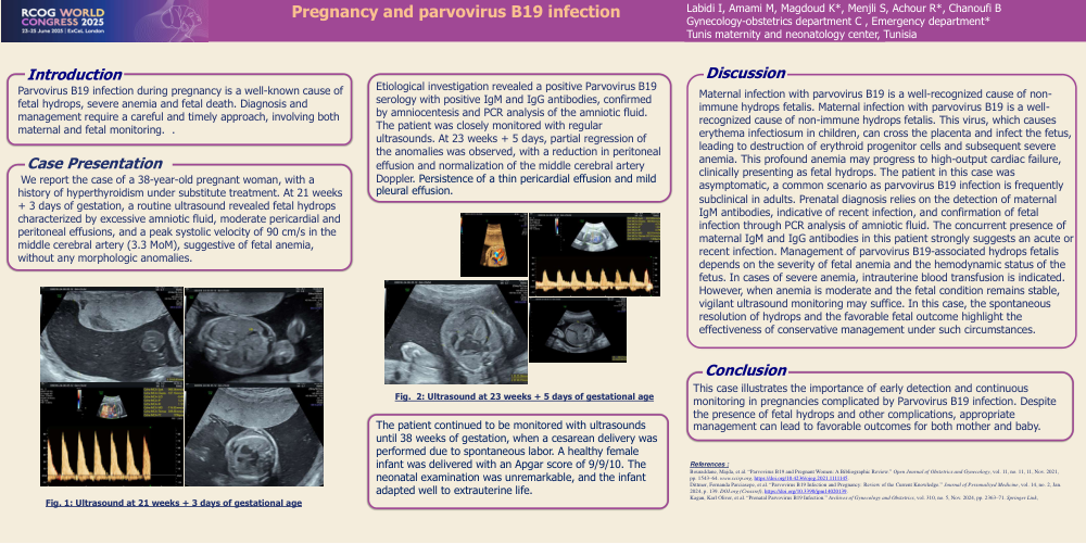
We report the case of a 38-year-old pregnant woman, with a history of hyperthyroidism under substitute treatment. At 21 weeks + 3 days of gestation, a routine ultrasound revealed fetal hydrops characterized by excessive amniotic fluid, moderate pericardial and peritoneal effusions, and a peak systolic velocity of 90 cm/s in the middle cerebral artery (3.3 MoM), suggestive of fetal anemia, without any morphologic anomalies.
Etiological investigation revealed a positive Parvovirus B19 serology with positive IgM and IgG antibodies, confirmed by amniocentesis and PCR analysis of the amniotic fluid. The patient was closely monitored with regular ultrasounds. At 23 weeks + 5 days, partial regression of the anomalies was observed, with a reduction in peritoneal effusion and normalization of the middle cerebral artery Doppler. Persistence of a thin pericardial effusion and mild pleural effusion.
The patient continued to be monitored with ultrasounds until 38 weeks of gestation, when a cesarean delivery was performed due to spontaneous labor. A healthy female infant was delivered with an Apgar score of 9/9/10. The neonatal examination was unremarkable, and the infant adapted well to extrauterine life.
Discussion:
Maternal infection with parvovirus B19 is a well-recognized cause of non immune hydrops fetalis.Maternal infection with parvovirus B19 is a well recognized cause of non-immune hydrops fetalis. This virus, which causes erythema infectiosum in children, can cross the placenta and infect the fetus, leading to destruction of erythroid progenitor cells and subsequent severe anemia. This profound anemia may progress to high-output cardiac failure, clinically presenting as fetal hydrops. The patient in this case was asymptomatic, a common scenario as parvovirus B19 infection is frequently subclinical in adults. Prenatal diagnosis relies on the detection of maternal IgM antibodies, indicative of recent infection, and confirmation of fetal infection through PCR analysis of amniotic fluid. The concurrent presence of maternal IgM and IgG antibodies in this patient strongly suggests an acute or recent infection. Management of parvovirus B19-associated hydrops fetalis depends on the severity of fetal anemia and the hemodynamic status of the fetus. In cases of severe anemia, intrauterine blood transfusion is indicated. However, when anemia is moderate and the fetal condition remains stable, vigilant ultrasound monitoring may suffice. In this case, the spontaneous resolution of hydrops and the favorable fetal outcome highlight the effectiveness of conservative management under such circumstances.
Conclusion:
This case illustrates the importance of early detection and continuous monitoring in pregnancies complicated by Parvovirus B19 infection. Despite the presence of fetal hydrops and other complications, appropriate management can lead to favorable outcomes for both mother and baby.