2,281 posters, 63 videos, 56 audios, 43 topics, 18 sessions, 6,174 authors, 2,323 institutions
ePostersLive by SciGen Technologies S.A. All rights reserved.
RCOG World Congress 2025
June 23 - 25, 2025 | ExCeL London, UK
EP.0049
QUANTITATIVE CHARACTERIZATION OF LUNG MICROSTRUCTURE BY ULTRASONIC BACKSCATTER SPECTROSCOPY
Poster Presenter
Authors
Yubia Amaya Guel, Juan M. Gallardo Gaona, Sandra Acevedo Gallegos, Ricardo Figueroa Damián, Mario Isaac Lumbreras Márquez, Diana Díaz Pérez, Iván Rosado Méndez, Irene Quiterio, Fabián Torres Robles, Raúl Esquivel Servent
Affiliations
Department of Maternal-Fetal Medicine. National Institute of Perinatology “Isidro Espinosa de Los Reyes”, Department of Pathological Anatomy. National Institute of Perinatology “Isidro Espinosa de Los Reyes”, Department of Medical Physics, University of Wisconsin - Madison, Institute of Physics, National Autonomous University of Mexico
Part of Topic
Advances in prenatal diagnosis
QUANTITATIVE CHARACTERIZATION OF LUNG MICROSTRUCTURE BY ULTRASONIC BACKSCATTER SPECTROSCOPY
Yubia Amaya-Guel a, Juan M. Gallardo-Gaona a, Sandra Acevedo-Gallegos a, Dulce M. Camarena a, Mario Isaac Lumbreras-Márquez a, Diana L. Díaz-Pérez b, Ivan Rosado-Méndez c, Irene G. Quiterio d, Fabian Torres-Robles d, R. Esquivel-Servent d
a Departamento de Medicina Materno Fetal. Instituto Nacional de Perinatología “Isidro Espinosa de Los Reyes”, Ciudad de México, México.
b Departamento de Anatomía Patológica. Instituto Nacional de Perinatología “Isidro Espinosa de Los Reyes”, Ciudad de México, México.
c Department of Medical Physics, University of Wisconsin - Madison, Madison, Wisconsin, United States of America.
d Instituto de Física, Universidad Nacional Autónoma de México, Ciudad de México, México.
Objectives: This project is a proof of concept and is part of the analysis of three different standardizations (sponge, animal and fetal), to propose the bases for the development of a new technique based on quantitative ultrasound, which has the capacity to determine fetal lung maturity.
Design: This was a cross-sectional study conducted in collaboration between the National Institute of Perinatalogy and the Institute of Physics of the UNAM
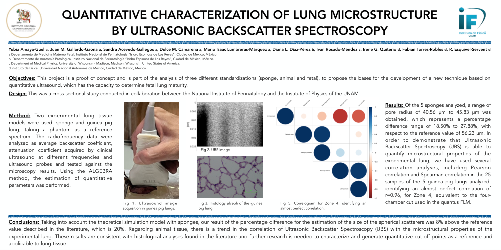
Method: Two experimental lung tissue models were used: sponge and guinea pig lung, taking a phantom as a reference spectrum. The radiofrequency data were analyzed as average backscatter coefficient, attenuation coefficient acquired by clinical ultrasound at different frequencies and ultrasound probes and tested against the microscopy results. Using the ALGEBRA method, the estimation of quantitative parameters was performed.
Results: Of the 5 sponges analyzed, a range of pore radius of 40.56 μm to 45.83 μm was obtained, which represents a percentage difference range of 18.50% to 27.88%, with respect to the reference value of 56.23 μm. In order to demonstrate that Ultrasonic Backscatter Spectroscopy (UBS) is able to quantify microstructural properties of the experimental lung, we have used several correlation analyses, including Pearson correlation and Spearman correlation in the 25 samples of the 5 guinea pig lungs analyzed, identifying an almost perfect correlation of r=0.96, for Zone 4, equivalent to the four-chamber cut used in the quantus FLM.
Conclusions: Taking into account the theoretical simulation model with sponges, our result of the percentage difference for the estimation of the size of the spherical scatterers was 8% above the reference value described in the literature, which is 20%. Regarding animal tissue, there is a trend in the correlation of Ultrasonic Backscatter Spectroscopy (UBS) with the microstructural properties of the experimental lung. These results are consistent with histological analyses found in the literature and further research is needed to characterize and generate quantitative cut-off points as a reference and applicable to lung tissue.