2,281 posters, 63 videos, 56 audios, 43 topics, 18 sessions, 6,174 authors, 2,323 institutions
ePostersLive by SciGen Technologies S.A. All rights reserved.
RCOG World Congress 2025
June 23 - 25, 2025 | ExCeL London, UK
EP.0398
Atypical Presentation of Hemoperitoneum Due to Ruptured Uterine Leiomyoma: A Case Report
Poster Presenter
Authors
Affiliations
Part of Topic
Endometriosis & uterine disorders
Atypical Presentation of Hemoperitoneum Due to Ruptured Uterine Leiomyoma: A Case Report
Doctor Zeinab Lami1, Doctor Rajaa Samaka2, Doctor Zahraa Lami3, Doctor Uzma Aziz1
1Tawam Hospital, Abu Dhabi, UAE. 2Ishtar Medical Center, Abu Dhabi, UAE. 3Latifa Hospital, Dubai, UAE
Objective:
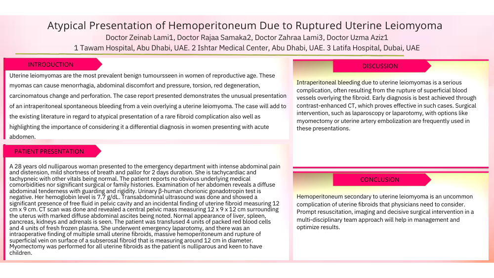
Uterine leiomyomas are the most prevalent benign tumoursseen in women of reproductive age. These myomas can cause menorrhagia, abdominal discomfort and pressure, torsion, red degeneration, carcinomatous change and perforation. The case report presented demonstrates the unusual presentation of an intraperitoneal spontaneous bleeding from a vein overlying a uterine leiomyoma. The case will add to the existing literature in regard to atypical presentation of a rare fibroid complication also well as highlighting the importance of considering it a differential diagnosis in women presenting with acute abdomen.
Case Report:
A 28 years old nulliparous woman presented to the emergency department with intense abdominal pain and distension, mild shortness of breath and pallor for 2 days duration. She is tachycardiac and tachypneic with other vitals being normal. The patient reports no obvious underlying medical comorbidities nor significant surgical or family histories. Examination of her abdomen reveals a diffuse abdominal tenderness with guarding and rigidity. Urinary β-human chorionic gonadotropin test is negative. Her hemoglobin level is 7.7 g/dL. Transabdominal ultrasound was done and showed a significant presence of free fluid in pelvic cavity and an incidental finding of uterine fibroid measuring 12 cm x 9 cm. CT scan was done and revealed a central pelvic mass measuring 12 x 9 x 12 cm surrounding the uterus with marked diffuse abdominal ascites being noted. Normal appearance of liver, spleen, pancreas, kidneys and adrenals is seen. The patient was transfused 4 units of packed red blood cells and 4 units of fresh frozen plasma. She underwent emergency laparotomy, and there was an intraoperative finding of multiple small uterine fibroids, massive hemoperitoneum and rupture of superficial vein on surface of a subserosal fibroid that is measuring around 12 cm in diameter. Myomectomy was performed for all uterine fibroids as the patient is nulliparous and keen to have children.
Discussion:
Intraperitoneal bleeding due to uterine leiomyomas is a serious complication, often resulting from the rupture of superficial blood vessels overlying the fibroid. Early diagnosis is best achieved through contrast-enhanced CT, which proves effective in such cases. Surgical intervention, such as laparoscopy or laparotomy, with options like myomectomy or uterine artery embolization are frequently used in these presentations.
Conclusions:
Hemoperitoneum secondary to uterine leiomyoma is an uncommon complication of uterine fibroids that physicians need to consider. Prompt resuscitation, imaging and decisive surgical intervention in a multi-disciplinary team approach will help in management and optimize results.