2,281 posters, 63 videos, 56 audios, 43 topics, 18 sessions, 6,174 authors, 2,323 institutions
ePostersLive by SciGen Technologies S.A. All rights reserved.
RCOG World Congress 2025
June 23 - 25, 2025 | ExCeL London, UK
EP.0384
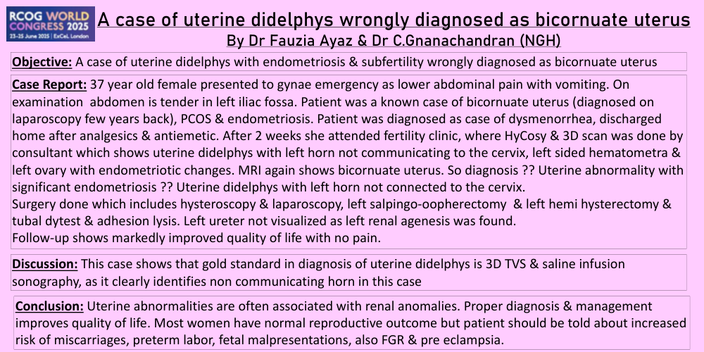
A case of uterine didelphys with endometriosis wrongly diagnosed as bicornuate uterus.
Poster Presenter
Authors
Affiliations
Part of Topic
Endometriosis & uterine disorders
Objective:A case of uterine didelphys with endometriosis & subfertility wrongly diagnosed as bicornuate uterus
Case Report: 37 year old female presented to gynae emergency as lower abdominal pain with vomiting. On examination abdomen is tender in left iliac fossa. Patient was a known case of bicornuate uterus (diagnosed on laparoscopy few years back), PCOS & endometriosis. Patient was diagnosed as case of dysmenorrhea, discharged home after analgesics & antiemetic. After 2 weeks she attended fertility clinic, where HyCosy & 3D scan was done by consultant which shows uterine didelphys with left horn not communicating to the cervix, left sided hematometra & left ovary with endometriotic changes. MRI again shows bicornuate uterus. So diagnosis ?? Uterine abnormality with significant endometriosis ?? Uterine didelphys with left horn not connected to the cervix.
Surgery done which includes hysteroscopy & laparoscopy, left salpingo-oopherectomy & left hemi hysterectomy & tubal dytest & adhesion lysis. Left ureter not visualized as left renal agenesis was found.
Follow-up shows markedly improved quality of life with no pain.
Discussion:This case shows that gold standard in diagnosis of uterine didelphys is 3D TVS & saline infusion sonography, as it clearly identifies non communicating horn in this case
Conclusion: Uterine abnormalities are often associated with renal anomalies. Proper diagnosis & management improves quality of life. Most women have normal reproductive outcome but patient should be told about increased risk of miscarriages, preterm labor, fetal malpresentations, also FGR & pre eclampsia.